Clinical Deep Dives
by Med School Audio - Medical Knowledge Reimagined & Learning Made Memorable.
Is this your podcast?Insights from recent episode analysis
Audience Interest
Podcast Focus
Publishing Consistency
Platform Reach
Insights are generated by CastFox AI using publicly available data, episode content, and proprietary models.
Total monthly reach
Estimated from 4 chart positions in 4 markets.
By chart position
- 🇮🇹IT · Medicine#1881K to 10K
- 🇰🇪KE · Medicine#630K to 100K
- 🇳🇬NG · Medicine#108500 to 3K
- 🇭🇰HK · Medicine#154500 to 3K
- Per-Episode Audience
Est. listeners per new episode within ~30 days
9.6K to 35K🎙 Daily cadence·538 episodes·Last published today - Monthly Reach
Unique listeners across all episodes (30 days)
32K to 116K🇰🇪86%🇮🇹9%🇳🇬3%+1 more - Active Followers
Loyal subscribers who consistently listen
18K to 64K
Market Insights
Platform Distribution
Reach across major podcast platforms, updated hourly
Total Followers
—
Total Plays
—
Total Reviews
—
* Data sourced directly from platform APIs and aggregated hourly across all major podcast directories.
On the show
Recent episodes
ANAHN 21: Vascular Supply of the Head and Neck - The Rivers That Sustain and Spread
May 12, 2026
49m 05s
ANAHN 20: Lymphatics of the Head and Neck - The Hidden Pathways of Disease and Defence
May 11, 2026
58m 45s
ANAHN 19: Anatomic Basis for Local Anesthesia - Mapping Silence in the Face
May 10, 2026
47m 56s
ANAHN 18: Cranial Nerves - The Twelve Messengers of the Mind
May 9, 2026
50m 17s
ANAHN 17: Brain and Spinal Cord - The Living Axis of Thought, Control, and Continuity
May 8, 2026
1h 13m 43s
Social Links & Contact
Official channels & resources
Official Website
Login
RSS Feed
Login
| Date | Episode | Description | Length | ||||||
|---|---|---|---|---|---|---|---|---|---|
| 5/12/26 | 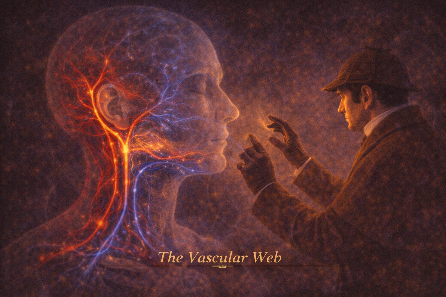 ANAHN 21: Vascular Supply of the Head and Neck - The Rivers That Sustain and Spread | This chapter is the circulatory map of the head and neck - a system of arteries that deliver, and veins that quietly return.But unlike a simple plumbing system, this network is:* Redundant* Interconnected* And clinically unforgivingBecause:* A blockage can blind* A rupture can flood* A connection can spread infection to the brainPART I - THE THREE GREAT SOURCESFrom the opening framework:Blood supply arises from:* External carotid artery* Internal carotid artery* Subclavian arteryConceptual ModelThink of it as:* External carotid → face and superficial structures* Internal carotid → brain and intracranial structures* Subclavian → neck, posterior structures, and indirect brain supplyThree rivers feeding one landscape - each with its own territory.PART II - COMMON CAROTID ARTERYOrigins* Right → brachiocephalic trunk* Left → aortic archKey Feature* No branches in the neck* Bifurcates at thyroid cartilage into:* External carotid* Internal carotidSpecial Structures* Carotid sinus → monitors blood pressure* Carotid body → monitors oxygen, CO₂, pHClinical Insight* Hypersensitivity → syncope with head movementAt the bifurcation, the body listens - measuring pressure, sensing life.PART III - EXTERNAL CAROTID ARTERYThe workhorse of the face and neckBranching Pattern* 6 collateral branches* 2 terminal branchesMajor Branches (Core Memory Set)1. Superior Thyroid* Supplies thyroid and larynx2. Ascending Pharyngeal* Supplies pharynx and skull base3. Lingual* Supplies tongue4. Facial* Supplies face5. Occipital* Supplies posterior scalp6. Posterior Auricular* Supplies ear regionTerminal Branches* Superficial temporal* MaxillaryPART IV - LINGUAL AND FACIAL ARTERIESLingual Artery* Runs deep to tongue* Supplies:* Tongue* Floor of mouth* Ends as deep lingual arteryClinical Note* Sublingual artery injury → surgical challengeFacial ArteryFrom page 339 diagram:* Tortuous path across face* Ends as angular artery near eyeKey Insight* Highly anastomotic → difficult to fully occlude bleedingThe face is never supplied by one vessel - it is a network of cooperation.PART V - MAXILLARY ARTERYThe deep supply of the faceThree Parts* Mandibular* Pterygoid* PterygopalatineKey Territories* Teeth (inferior alveolar artery)* Muscles of mastication* Nasal cavity* PalateClinical Insight* Middle meningeal artery → risk in skull fractures* Dental procedures → bleeding riskPART VI - INTERNAL CAROTID ARTERYKey Rule* No branches in the neckFunction* Supplies:* Brain* Orbit* ForeheadMajor Contribution* Ophthalmic arteryClinical Insight* Central retinal artery blockage → sudden blindnessWhen this vessel fails - vision and consciousness are at stake.PART VII - SUBCLAVIAN ARTERYThree Parts (relative to anterior scalene)* Medial* Posterior* LateralKey Branches* Vertebral artery → brain* Thyrocervical trunk → neck structures* Costocervical trunk* Dorsal scapularKey Concept* Provides collateral circulation with carotid systemPART VIII - VENOUS DRAINAGETwo Main Systems* Internal jugular vein* External jugular veinPART IX - FACIAL VEINS AND DANGERFacial Vein* Connects:* Face* Orbit* Cavernous sinusCritical Feature* No valves → bidirectional flowClinical Danger ZoneTriangle:* Nose* Upper lip* Medial eyeRisk* Infection → cavernous sinus thrombosisWhat begins as a small infection - can travel inward to the brain.PART X - PTERYGOID VENOUS PLEXUSFrom page 351 diagram:* Dense venous network in deep face* Communicates with:* Cavernous sinus* Nasal cavity* OrbitClinical Insight* Dental injections → risk of haematoma or spread of infectionPART XI - INTERNAL JUGULAR VEINMain Drain* Brain* Face* NeckTributaries* Facial vein* Lingual vein* Thyroid veinsPath* Jugular foramen → brachiocephalic veinPART XII - EXTERNAL JUGULAR VEIN* Superficial* Formed by:* Posterior auricular* Retromandibular veinsClinical Use* Visible marker of venous pressureInsight* Engorgement → right heart failurePART XIII - FINAL INTEGRATIONArterial System* Delivers oxygen* Highly branched* RedundantVenous System* Drains blood* Highly interconnected* Potential route for diseaseArteries nourish.Veins reveal.Key Takeaways* Three arterial sources: carotid (internal/external) and subclavian* External carotid supplies face and neck* Internal carotid supplies brain and orbit* Maxillary artery is key for deep face* Venous system lacks valves → allows spread of infection* Facial vein connects to cavernous sinus (danger zone)* Internal jugular is the main venous drainage* External jugular reflects systemic venous pressure This is a public episode. If you'd like to discuss this with other subscribers or get access to bonus episodes, visit drmanaankarray.substack.com/subscribe | 49m 05s | ||||||
| 5/11/26 | 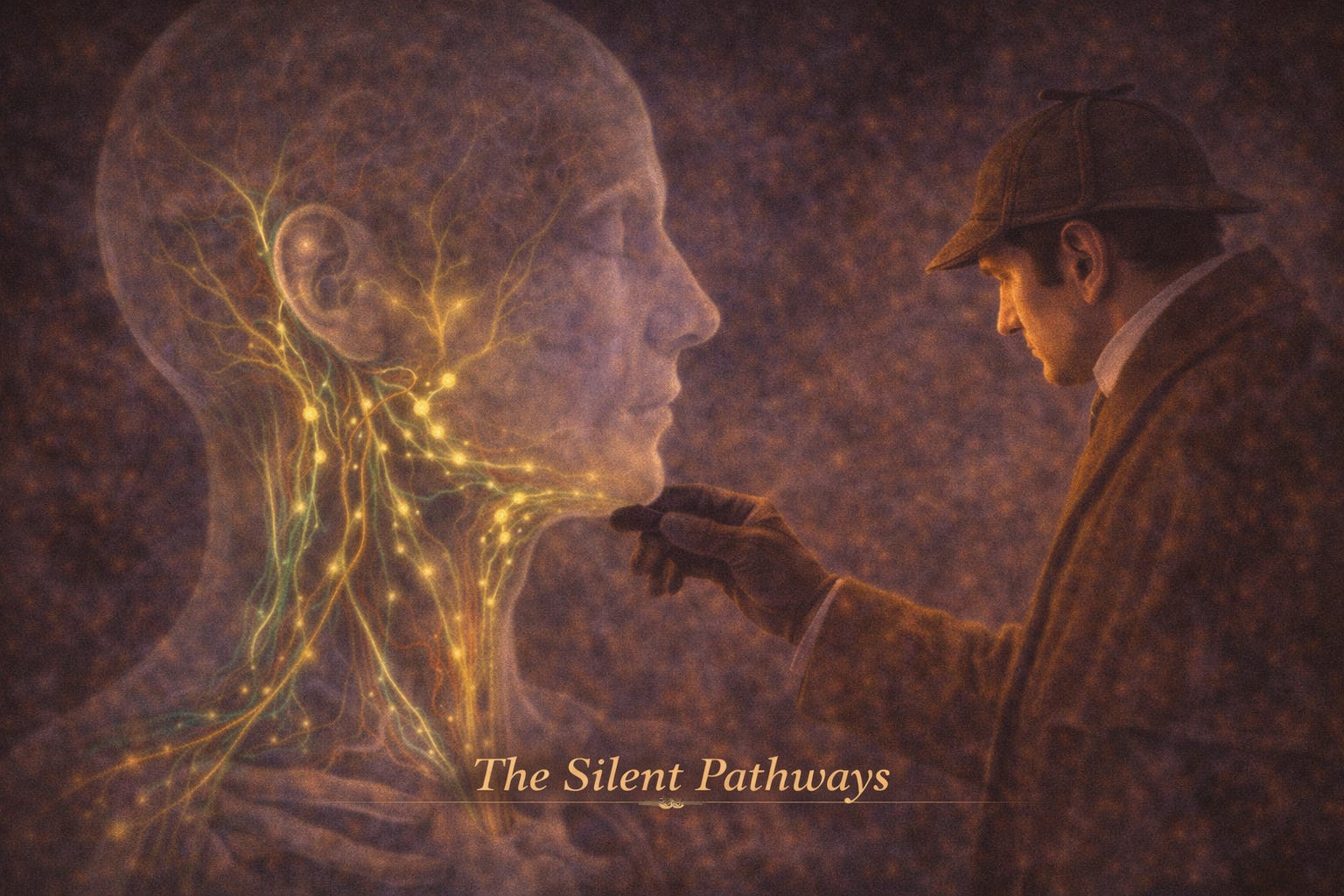 ANAHN 20: Lymphatics of the Head and Neck - The Hidden Pathways of Disease and Defence | If Chapter 19 was about interrupting sensation,this chapter is about something quieter - and arguably more powerful:tracking disease through the body.Because the lymphatic system does not shout.It signals.It tells you:* Where infection started* Where cancer may spread* Where the body is fighting backAnd it does this through:* Nodes* Channels* PatternsPART I - WHAT IS LYMPH?From the opening section:Lymph is:* Extracellular fluid from interstitial spaces* Derived from capillaries* Unable to re-enter veins directly due to pressure differencesWhat does it carry?* Proteins* Fats* Cells* DebrisWhere does it go?* Small lymphatic vessels → larger vessels* Eventually drains into:* Right lymphatic duct* Thoracic duct* Then into subclavian veinsBlood circulates.Lymph returns what is left behind.PART II - LYMPH NODES: THE FILTERING STATIONSDefined as:* Structures that filter lymph* Sites of immune activityKey ConceptLymph passes through:* At least one node* Usually severalWhat happens inside?* Foreign material → phagocytosed* Immune response activatedEvery node is a checkpoint - where the body asks: friend or threat?PART III - LYMPH NODES OF THE HEADFrom page 328:Important rule:* No lymph nodes inside the brain* All are extracranialMajor GroupsOccipital* Back of scalpMastoid (postauricular)* Behind earPreauricular* In front of earParotid* Around parotid glandWhat they doDrain:* Scalp* Ear* Superficial facePART IV - LYMPH NODES OF THE FACEFrom page 328:Three key systems:1. Superficial Facial Nodes* Along facial vessels* Includes:* Infraorbital* Buccal* Mandibular2. Deep Facial Nodes* Along maxillary artery* In infratemporal region3. Special Groups* Lingual nodes → tongue* Retropharyngeal nodes → behind pharynxThe face drains inward - toward deeper, less visible systems.PART V - LYMPH NODES OF THE NECKFrom pages 328–329:Superficial System* Submental* Submandibular* Superficial cervicalDeep System (Critical)* Deep cervical chain along internal jugular vein* Divided into:* Superior deep cervical* Inferior deep cervicalThe Final PathAll lymph ultimately reaches:* Deep cervical nodes* Then → jugular trunk → venous systemEverything flows downward - toward a final convergence.PART VI - THE KEY NODES (CLINICAL LANDMARKS)Jugulodigastric NodeFrom page 332:* Receives lymph from:* Tonsils* Tongue* Easily palpableWhy it matters* First sign of oral disease* Called:* “Tonsillar node”* “Sentinel node”Jugulo-omohyoid Node* Drains tongue* Located lower in neckSome nodes are not just filters - they are signals of deeper pathology.PART VII - THE MAP OF DRAINAGEFrom the diagram on page 329:You can visualise:* Green lymphatic channels* Flow from face → neck → deep chainCore Principle* Superficial → deep* Regional → centralPART VIII - DRAINAGE OF SPECIFIC STRUCTURESFrom pages 331–333:Face* Drains to submandibular nodesTongue (Complex)Three systems:* Tip → submental* Lateral anterior → jugulodigastric* Central → jugulo-omohyoidTeeth* Incisors → submental* Others → submandibularPharynx & Sinuses* Retropharyngeal nodes* Deep cervical chainThe tongue does not drain symmetrically - it crosses sides, blurring boundaries.PART IX - CLINICAL THREADS1. Lymph Node EnlargementFrom page 333:Nodes become:* Swollen* Hard* PainfulIndicates:* Infection* Inflammation* Cancer2. Disease Mapping* Location of enlarged node → site of disease3. Primary vs Secondary Nodes* Primary node = first barrier* Secondary = next stage4. Cancer Spread* Travels via lymphatics* May require block dissection surgeryThe lymphatic system does not prevent spread - it slows and reveals it.PART X - CLINICAL EXAMINATIONKey nodes to examine:* Submental* Submandibular* Deep cervical chainTechnique:* Palpate along sternocleidomastoid* Assess:* Size* Tenderness* ConsistencyA skilled hand can read diseasebefore imaging ever sees it.Key Takeaways* Lymph collects excess interstitial fluid* Lymph nodes filter and respond to pathogens* Head and neck drainage follows predictable pathways* Deep cervical nodes are the final common pathway* Jugulodigastric node is clinically critical* Lymphatic mapping helps localise disease* Node examination is a key diagnostic tool This is a public episode. If you'd like to discuss this with other subscribers or get access to bonus episodes, visit drmanaankarray.substack.com/subscribe | 58m 45s | ||||||
| 5/10/26 | 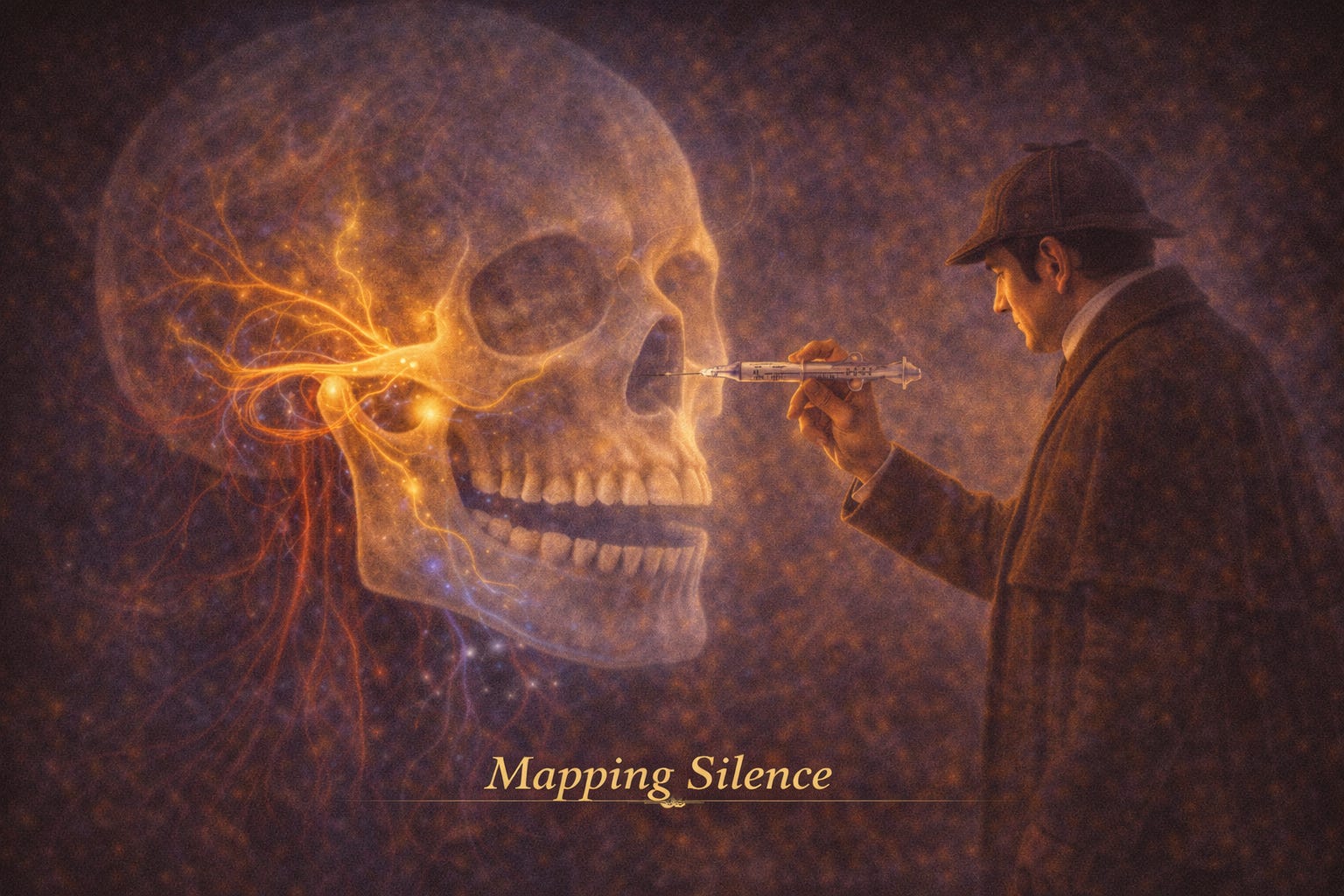 ANAHN 19: Anatomic Basis for Local Anesthesia - Mapping Silence in the Face | If Chapter 18 gave us the wiring of the cranial nerves,this chapter teaches us something far more practical:How to interrupt that wiring - safely, deliberately, and effectively.This is not just anatomy.This is applied anatomy - where knowledge becomes intervention.PART I - WHAT IS ANAESTHESIA, REALLY?From page 312:* Anaesthesia = loss of sensation due to drugs, injury, or diseaseMechanismLocal anaesthetics:* Stabilise nerve membranes* Block conduction of impulses* Prevent transmission of sensationFibre Sensitivity (Clinical Gold)Order of blockade:* Pain fibres (small, unmyelinated) → first* Touch/proprioception → later* Motor → lastPain disappears first - because it travels along the most fragile pathways.PART II - TWO STRATEGIES: INFILTRATION VS BLOCK1. Infiltration (Local)* Inject near nerve endings* Small, localised effect2. Nerve Block (Trunk Anaesthesia)* Inject near nerve trunk* Large region anaesthetisedA Third Concept: Plexus Anaesthesia* Injection into connective tissue over periosteum* Relies on diffusion through bone* Works best where bone is thin (maxilla)The difference is simple:* Infiltration whispers* Plexus spreads* Blocks silence entire conversationsPART III - THE MAXILLA: WHERE DIFFUSION WORKSFrom pages 312–314:Maxillary bone:* Thin cortical plate* Allows anaesthetic diffusionNerve Supply* Anterior superior alveolar* Middle superior alveolar* Posterior superior alveolarKey Insight* Plexus anaesthesia is ideal in maxilla* Especially effective except around first molar regionClinical Image (Page 315)The diagram shows:* Needle placed near premolar apex* Pink-highlighted area showing spread across teethThis visually reinforces:Diffusion-based anaesthesia works when anatomy allows it.PART IV - THE MANDIBLE: WHERE DIFFUSION FAILSFrom page 316:Mandibular bone:* Thick cortical plate* Prevents diffusionConsequence* Plexus anaesthesia limited to incisors* Trunk (nerve block) requiredThe mandible teaches a hard lesson:when structure resists, strategy must change.PART V - MAXILLARY NERVE BLOCKS (THE PRECISION MAP)Posterior Superior Alveolar (PSA) BlockFrom page 317:* Anaesthetises molars* But may miss mesial root of first molar (~28%)Clinical risk:* Nearby artery → hematoma (page 318 image)Infraorbital BlockFrom page 318–319:* Covers incisors → canine (and often premolars)* Access via infraorbital foramenCritical warning:* Too deep → orbital complications (eye muscle paralysis)Palatal BlocksGreater Palatine* Posterior hard palateNasopalatine* Anterior palateFrom pages 320–321:* Nasopalatine block anaesthetises both sides* Painful due to tightly bound mucosaThe palate is not forgiving - it demands slow, deliberate technique.PART VI - MANDIBULAR NERVE BLOCKS (THE CORE SYSTEM)Inferior Alveolar Nerve BlockFrom page 322:* Target: mandibular foramen* Anaesthetises:* Teeth* Gingiva* Often lingual nerve as wellKey Landmarks* Retromolar pad* Pterygomandibular foldClinical RealityFrom page 323:* Failure rate: 15–20%* Positive aspiration: 10–15% (highest)This is not a simple injection - it is navigation through variable anatomy.PART VII - SUPPLEMENTARY BLOCKSBuccal Nerve Block* Buccal gingiva of molarsMental Nerve Block* Lower lip, chin, anterior gingivaIncisive Nerve Block* Pulp of anterior teethFrom pages 323–326:* Mental and incisive nerves = terminal branches of inferior alveolar nerveClinical Insight* Mental block → soft tissue* Incisive block → pulpal anaesthesiaPART VIII - THE MOST IMPORTANT SAFETY STEPAspirationFrom page 311 & 314:* Pull back syringe before injecting* If blood appears → DO NOT injectWhy it matters* Intravascular injection → toxicity* Can affect:* Heart* Brain* Local tissueThis is the moment of pause - where precision becomes safety.PART IX - WHAT THE TABLES SHOW (PAGE 313)The tables map:* Which block → which tooth* Pulp vs gingiva vs palateKey takeaway:* No single technique covers everything* Combination strategies are often requiredAnaesthesia is not a single act - it is a carefully choreographed sequence.Key Takeaways* Local anaesthesia blocks nerve conduction* Pain fibres are blocked first* Maxilla → diffusion works (plexus)* Mandible → requires nerve blocks* Each block targets a specific anatomical pathway* Aspiration is critical for safety* Clinical success depends on anatomy + technique This is a public episode. If you'd like to discuss this with other subscribers or get access to bonus episodes, visit drmanaankarray.substack.com/subscribe | 47m 56s | ||||||
| 5/9/26 | 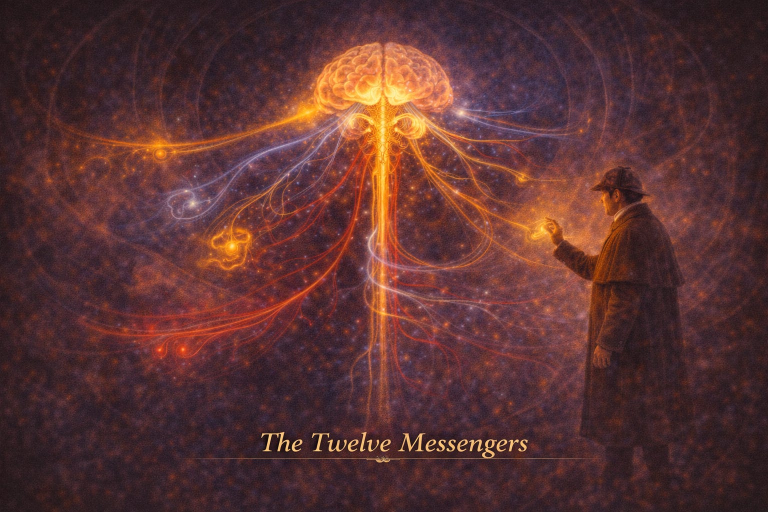 ANAHN 18: Cranial Nerves - The Twelve Messengers of the Mind | If Chapter 17 was the axis,this chapter is the distribution network.From the brainstem emerge twelve distinct pathways - each with:* A purpose* A territory* A vulnerabilityTogether, they transform central command into lived experience:* Sight* Sound* Expression* Swallowing* SpeechPART I - THE GRAND DESIGN: 12 CRANIAL NERVESFrom the opening section:* 12 paired nerves arise from the brain and exit via skull foraminaThe Sequence (Rostral → Caudal)I. OlfactoryII. OpticIII. OculomotorIV. TrochlearV. TrigeminalVI. AbducensVII. FacialVIII. VestibulocochlearIX. GlossopharyngealX. VagusXI. AccessoryXII. HypoglossalThink of them as twelve emissaries - each carrying a distinct dialect of the nervous system.PART II - THE LANGUAGE OF NERVES (MODALITIES)From pages 278–279:Cranial nerves do not all speak the same “language” - they carry specific modalitiesMotor Modalities* GSE → skeletal muscle* GVE → smooth muscle, glands (parasympathetic)* SVE → branchial arch musclesSensory Modalities* GSA → touch, pain, temperature* GVA → visceral sensation* SSA → vision, hearing* SVA → taste, smellEach nerve is not just a wire - it is a bundle of meanings.PART III - THE SENSORY GATEWAYSI. Olfactory (Smell)* Pure SVA* From nasal mucosa → olfactory bulbClinical: anosmiaII. Optic (Vision)* Pure SSA* Retina → optic chiasm → brainFrom page 283 diagram:* Partial crossing at chiasm explains visual field defectsSmell and sight do not pass through relay stations - they go directly to perception.PART IV - THE EYE MOVERSIII, IV, VI → Control eye movementIII. Oculomotor* Most eye muscles* Parasympathetic → pupil constrictionClinical:* “Down and out” eye* Dilated pupilIV. Trochlear* Superior obliqueClinical:* Vertical diplopiaVI. Abducens* Lateral rectusClinical:* Eye deviates mediallyThree nerves, one purpose:to align perception with reality.PART V - THE TRIGEMINAL: THE GREAT SENSORV. TrigeminalFrom pages 285–296:* Largest cranial nerve* Sensory to face* Motor to masticationThree DivisionsV1 (Ophthalmic)* Sensory only* Forehead, eye, noseV2 (Maxillary)* Sensory only* Midface, upper teethV3 (Mandibular)* Mixed* Lower face + chewing musclesClinical:* Trigeminal neuralgia → severe facial painIf the face could speak,it would speak through V.PART VI - THE FACE AND EXPRESSIONVII. Facial NerveFrom pages 299–302:Carries nearly every modality:* Motor → facial expression* Taste → anterior 2/3 tongue* Parasympathetic → glands* Sensory → earClinical: Bell’s palsy* Facial droop* Loss of expressionThis is the nerve of identity - it turns feeling into visible emotion.PART VII - HEARING AND BALANCEVIII. Vestibulocochlear* Cochlear → hearing* Vestibular → balanceClinical (page 303):* Ménière disease → vertigo, tinnitus* Conductive vs nerve deafnessIt does not just hear the world - it tells you where you are within it.PART VIII - THE THROAT AND VISCERAIX. Glossopharyngeal* Taste posterior 1/3* Parotid secretion* Swallowing* Carotid body/sinusX. VagusFrom pages 305–307:* Most extensive nerve* Controls:* Heart* Lungs* Gut* VoiceClinical:* Damage → swallowing, speech, life-threatening issuesThe vagus is not a nerve - it is a bridge between mind and body.PART IX - POSTURE AND TONGUEXI. Accessory* Sternocleidomastoid* TrapeziusClinical:* Shoulder droopXII. Hypoglossal* Motor to tongueClinical:* Tongue deviates toward lesionEven speech depends on alignment - of muscle, nerve, and intention.PART X - CLINICAL TESTING (THE EXAM MAP)From Table 18-5 (pages 293–294):Each nerve can be tested through:* Movement* Sensation* ReflexExamples:* CN II → visual fields* CN V → facial sensation* CN VII → facial symmetry* CN IX/X → gag reflexThe cranial nerve exam is not a checklist - it is a conversation with the brain through the body.Key Takeaways* 12 cranial nerves = functional pathways from brain* Each nerve carries specific modalities* Some are pure (I, II, VIII)* Some are mixed (V, VII, IX, X)* Clinical testing localises lesions precisely* Integration across nerves enables complex behaviours This is a public episode. If you'd like to discuss this with other subscribers or get access to bonus episodes, visit drmanaankarray.substack.com/subscribe | 50m 17s | ||||||
| 5/8/26 | 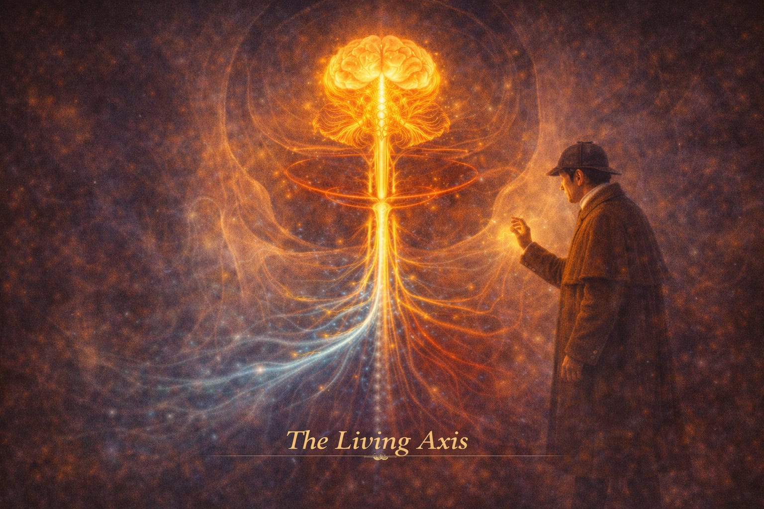 ANAHN 17: Brain and Spinal Cord - The Living Axis of Thought, Control, and Continuity | If Chapter 16 was the gateway,this chapter is the command centre and the highway combined.Here, structure becomes function:* Protection becomes layered* Fluid becomes cushioning intelligence* Tissue becomes thoughtAnd at its core:* The brain interprets* The spinal cord conducts* The system sustains lifePART I - THE CENTRAL NERVOUS SYSTEMFrom the opening page:The brain and spinal cord form the central nervous system (CNS) - the body’s integrating and responding systemIf the body is a city,this is both government and infrastructure.PART II - MENINGES: THE PROTECTIVE ENVELOPEThree layers surround the CNS:* Dura mater → tough outer layer* Arachnoid mater → web-like middle layer* Pia mater → delicate layer adhering to brain surfaceFunctional InsightFrom page 264:* Subarachnoid space contains CSF and blood vessels* Arachnoid granulations → drain CSF into venous sinusesProtection here is not rigid - it is layered, fluid, and dynamic.PART III - CEREBROSPINAL FLUID: THE SILENT CUSHION* Produced by choroid plexus* Circulates through ventricles → subarachnoid space* Reabsorbed into venous systemFunctions* Shock absorption* Nutrient transport* Pressure bufferingThe brain does not sit - it floats.PART IV - THE BRAIN: THREE GRAND DIVISIONSAlthough embryology begins with five parts, the adult brain shows three dominant regions1. Cerebral HemispheresFrom pages 265–267:* Largest component* Responsible for:* Sensation* Memory* Learning* Voluntary movementKey Features* Gyri (ridges) and sulci (grooves)* Lobes:* Frontal → motor, planning* Parietal → sensory* Temporal → hearing* Occipital → vision* Insula → tasteFrom the lateral brain diagram (page 266):* Central sulcus separates motor and sensory cortex* Temporal lobe sits like a “thumb” inferiorlyThe cortex is a landscape - each fold a compressed story of function.2. Cerebellum* Coordinates movement* Maintains balance* Refines motor activityFunctional divisions:* Neocerebellum → precision* Paleocerebellum → posture* Archicerebellum → spatial orientationIt does not initiate movement - it perfects it.3. BrainstemFrom page 269 onward:* Controls vital functions:* Breathing* Heart rate* Blood pressure* Origin of most cranial nervesPART V - THE BRAINSTEM: THE LIFE COREDiencephalon* Thalamus → sensory relay* Hypothalamus → homeostasis, endocrine control* Epithalamus → pineal gland* Subthalamus → motor integrationMesencephalon (Midbrain)* Visual + auditory reflexes* Superior & inferior colliculiMetencephalon* Pons (visible bulge)* Cerebellar connections* Cranial nerves V–VIIIMyelencephalon (Medulla)From page 272:* Contains vital centres for life* Pyramidal decussation → crossing of motor fibres* Cranial nerves IX–XIIDamage here is not deficit - it is catastrophe.PART VI - VENTRICULAR SYSTEMFour ventricles:* Two lateral* Third* FourthFlow PathCSF travels:* Lateral → Third → Aqueduct → Fourth → Subarachnoid spaceFrom page 272–273:* Exit via:* Foramen of Magendie* Foramina of LuschkaPART VII - BLOOD SUPPLY: THE CIRCLE OF LIFEFrom page 273–274:Two main sources:* Internal carotid arteries* Vertebral arteries → form basilar arteryCircle of WillisFrom the diagram (page 274):* Anterior cerebral* Middle cerebral* Posterior cerebral* Communicating arteriesClinical Insight* Poor collateral supply in deeper brain* Occlusion → permanent damageBlood here is not just supply - it is permission to function.PART VIII - VENOUS DRAINAGE* Pial venous plexus → cerebral veins* Drain into venous sinuses* Deep structures → great cerebral vein → straight sinusPART IX - THE SPINAL CORD: THE HIGHWAYDefined as:* Continuation of medulla* Ends at L1–L2 (conus medullaris)Key Structures* Filum terminale → anchors cord* Cauda equina → nerve roots below cordCross-Section InsightFrom page 277 diagram:* Outer white matter → tracts* Inner grey matter → H-shapedFunctional Layout* Dorsal horns → sensory* Ventral horns → motor* Lateral horn (T1–L2) → sympathetic outputThe spinal cord is not passive - it is a decision-maker in motion.PART X - REFLEXES: THE RAPID CIRCUITFrom the reflex diagram (page 277):Two systems:* Somatic reflex → skeletal muscle* Visceral reflex → autonomic responseKey idea:* Reflexes bypass higher centres → speedPART XI - CLINICAL THREADSStroke* Caused by arterial occlusion* Leads to neurological deficitsBrainstem Damage* Often fatal due to vital centresLumbar Puncture* Performed in lumbar cistern* Safe due to cauda equina mobilityKey Takeaways* CNS = brain + spinal cord* Meninges + CSF protect and cushion* Brain has three main divisions* Brainstem controls vital functions* Blood supply is critical and vulnerable* Spinal cord transmits and processes signals* Reflexes enable rapid responses This is a public episode. If you'd like to discuss this with other subscribers or get access to bonus episodes, visit drmanaankarray.substack.com/subscribe | 1h 13m 43s | ||||||
| 5/7/26 |  ANAHN 16: Palate, Pharynx, and Larynx - The Gateways of Breath, Voice, and Passage | If Chapter 15 was the living floor,this chapter is the gateway system.Three overlapping purposes unfold here:* Separation (air vs food)* Protection (airway vs aspiration)* Expression (voice vs silence)And at the centre of it all:* The palate closes* The pharynx channels* The larynx speaksPART I - THE PALATE: THE FIRST GATEFrom the opening section:The palate forms the roof of the mouth and floor of the nasal cavityTwo Distinct WorldsHard Palate - The Fixed Platform* Bony (maxilla + palatine bones)* Immovable* Provides surface for tongue to press foodFrom page 244–245:* Covered by specialised mucosa* Divided into regions (median raphe, glandular areas)Soft Palate - The Mobile Gate* Muscular* Suspended* Ends in the uvulaKey function:* Seals the nasopharynx during swallowingMuscles of the Soft PalateFrom Table 16-1:* Levator veli palatini → elevates* Tensor veli palatini → tenses* Musculus uvulae → lifts uvula* Palatoglossus → closes fauces* Palatopharyngeus → elevates pharynxInnervation Rule* Mostly via vagus nerve (pharyngeal plexus)* Exception → Tensor veli palatini (CN V3)The palate is not just a roof.It is a door that closes at exactly the right moment.PART II - THE PHARYNGEAL PLEXUS: THE CONTROL NETWORKA key concept early in the chapter:* Sensory → Glossopharyngeal (CN IX)* Motor → Vagus (CN X)* Vasomotor → Sympathetic fibresThink of this as a neural parliament - multiple voices, one coordinated decision.PART III - THE PHARYNX: THE SHARED PASSAGEDefined as:* A fibromuscular tube from skull base → oesophagusLength:* ~12–14 cmThree Regions1. Nasopharynx* Behind nose* Contains:* Pharyngeal tonsil (adenoids)* Opening of auditory tubeKey function:* Air passage2. Oropharynx* From soft palate → epiglottis* Contains palatine tonsilsKey function:* Air + food3. Laryngopharynx* From epiglottis → oesophagus* Leads to:* Larynx (air)* Oesophagus (food)This is the crossroads of life - where direction determines survival.PART IV - THE PHARYNGEAL WALL: THE MOVING TUBEThree layers:* Mucosa* Fibromuscular layer* Outer fasciaConstrictor MusclesFrom page 253 and Fig 16-3:* Superior* Middle* InferiorThey:* Overlap like telescoping sleeves* Push food downwardKey Functional Insight* Inferior constrictor → forms upper oesophageal sphincterLongitudinal Muscles* Stylopharyngeus (only one innervated by CN IX)* Salpingopharyngeus* PalatopharyngeusThey:* Elevate pharynx and larynxPART V - THE TONSILS: THE GUARDIANSPart of Waldeyer’s ring:* Palatine tonsil* Pharyngeal tonsil* Lingual tonsilKey Role* Immune defence at entry pointClinical InsightFrom page 249:* Tonsillectomy risk:* Close to carotid artery* Rich vascular supply* Glossopharyngeal nerve nearbyPART VI - THE OESOPHAGUS: THE DESCENT* Begins at C6 (cricoid level)* Lies behind trachea* Recurrent laryngeal nerve in grooveMuscle Transition* Upper → skeletal* Lower → smooth* Middle → mixedPART VII - THE LARYNX: THE VOICE AND VALVEDefined as:* Air passage* Protective sphincter* Organ of phonationKey StructuresCartilages (9 total)* Unpaired:* Thyroid* Cricoid* Epiglottis* Paired:* Arytenoid* Corniculate* CuneiformVocal ApparatusFrom Fig 16-7:* False cords (ventricular folds)* True cords (vocal folds)Movements* Adduction → close cords* Abduction → open cordsSound:* Air passes → vibration → voiceCritical Muscle* Posterior cricoarytenoid→ ONLY muscle that opens airwayPART VIII - NERVE SUPPLY OF LARYNXAll from vagus nerve:* Superior laryngeal:* Internal → sensory above cords* External → motor (cricothyroid)* Recurrent laryngeal:* Motor to all other muscles* Sensory below cordsPART IX - DEGLUTITION: THE ORCHESTRATED ACTFrom page 262:Stage 1 - Voluntary* Tongue pushes bolusStage 2 - Involuntary* Soft palate elevates → seals nose* Pharynx preparesStage 3 - Final* Constrictors push bolus into oesophagusProtective Mechanisms* Epiglottis redirects* Vocal cords close* Aryepiglottic folds form a chuteSwallowing is not a single act.It is a precise choreography where failure is dangerous.PART X - CLINICAL THREADSAdenoids* Enlarged pharyngeal tonsil* Causes mouth breathing, snoringPiriform Recess* Site where fish bones lodge* Internal laryngeal nerve at riskRecurrent Laryngeal Nerve Injury* Hoarseness → voice loss → airway compromiseAirway Emergencies* Heimlich manoeuvre* Cricothyrotomy (emergency airway)Key Takeaways* Palate separates nasal and oral cavities* Pharynx is a shared pathway for air and food* Constrictors drive swallowing* Larynx protects airway and produces voice* Vagus nerve dominates control* Deglutition is a coordinated, multi-phase reflex This is a public episode. If you'd like to discuss this with other subscribers or get access to bonus episodes, visit drmanaankarray.substack.com/subscribe | 51m 16s | ||||||
| 5/6/26 | 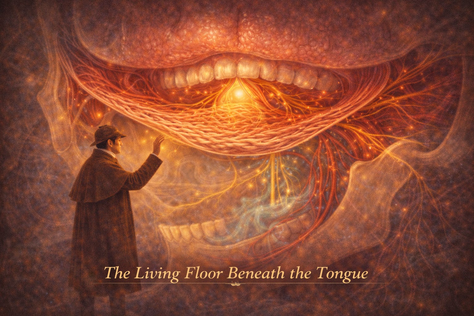 ANAHN 15: Submandibular Region and Floor of Mouth - The Living Foundation of Speech and Swallow | If the previous episode was about hidden corridors,this chapter is about living ground.Because here, beneath the tongue, lies a region that:* Lifts* Moves* Secretes* CoordinatesIt is not static anatomy.It is functional architecture in motion.And everything converges here:* Air becomes speech* Food becomes swallow* Thought becomes articulationPART I - THE SUBMANDIBULAR REGION: THE FOUNDATIONDefined as the space between:* Mandible (above)* Hyoid bone (below)This is a transition zone:* Between head and neck* Between structure and functionContained within:* Suprahyoid muscles* Tongue musculature* Submandibular and sublingual glandsBoundaries (Think: The Triangle)From the description on page 230:* Superior → Inferior border of mandible* Inferolateral → Anterior & posterior bellies of digastric* Floor → Mylohyoid muscleA triangle that supports the tongue above it - like a sling.PART II - MUSCLES OF THE FLOOR: THE SUSPENSION SYSTEMFrom the table on page 231 (Table 15-1), the key players:Suprahyoid Muscles* Digastric* Stylohyoid* Mylohyoid* GeniohyoidCore ConceptAll attach to the hyoid bone.And together they:* Elevate the floor of the mouth* Assist swallowing* Help open the jawMylohyoid - The True FloorFrom the diagram on page 232 (Fig 15-1):* Forms a muscular sheet* Meets its partner at the midline (median raphe)* Supports the tongue above itThis is the “floorboard” of the oral cavity.Digastric - The Dual ForceTwo bellies:* Anterior → pulls hyoid forward* Posterior → pulls hyoid backwardTogether:* Elevate hyoid* Open the mouth when hyoid is fixedA muscle of balance - pulling in two directions to create control.PART III - THE TONGUE: SHAPE AND DIRECTIONThe tongue is not a single muscle.It is a muscular orchestra.Two SystemsFrom page 233–235:1. Intrinsic Muscles* Longitudinal* Transverse* VerticalFunction:* Change shape of tongue2. Extrinsic Muscles* Genioglossus → protrudes* Hyoglossus → depresses* Styloglossus → retracts* Palatoglossus → elevates posterior tongueFunction:* Control direction of movementFrom the diagram on page 234 (Fig 15-4):* You can see fibres fanning, crossing, interminglingNo single movement is isolated.Every action is coordinated complexity.Innervation Rule* All tongue muscles → Hypoglossal nerve (CN XII)* Exception → Palatoglossus (pharyngeal plexus)PART IV - SALIVARY GLANDS: THE MOISTURE SYSTEMTwo major glands live here:* Submandibular gland* Sublingual glandSubmandibular GlandFrom page 238 and Fig 15-9:* Located in submandibular triangle* Extends into floor of mouth* Drains via Wharton’s duct → sublingual caruncleSublingual Gland* Lies beneath tongue* Above mylohyoid* Drains via multiple small ducts (Rivinus)* Sometimes forms a larger duct (Bartholin)These glands are quiet workers - ensuring lubrication, digestion, and speech.Innervation (The Secretory Pathway)From page 239:* Parasympathetic → Facial nerve (via chorda tympani)* Synapse → Submandibular ganglion* Travel via → Lingual nerve (V3)A beautiful relay:Facial nerve → Lingual nerve → GlandsPART V - NERVES: THE COMMUNICATION NETWORKTrigeminal Nerve (V3)* Lingual nerve:* General sensation to anterior 2/3 of tongue* Carries taste (via chorda tympani)Hypoglossal Nerve (CN XII)* Motor to tongue* Runs deep across carotid system* Ends at tongue tipFrom the diagram on page 240 (Fig 15-10):* You can trace its course beneath muscles toward the tonguePART VI - BLOOD SUPPLY: THE FLOWLingual Artery* Branch of external carotid* Supplies tongue and floorKey branches:* Deep lingual* Sublingual* Dorsal lingualFacial Artery* Supplies submandibular gland* Gives submental branchVenous Drainage* Deep lingual veins* Drain into:* Facial vein* Internal jugular veinPART VII - LYMPHATIC DRAINAGE: THE HIDDEN EXITFrom page 242:* Submandibular nodes drain:* Lips* Nose* Tongue* Key node:* Jugulodigastric node (principal node of tongue)This is where disease travels quietly before it is seen.PART VIII - CLINICAL THREADS1. Tongue Cancer* Most common oral cavity cancer* Often squamous cell carcinoma* Early spread to deep cervical nodes2. Hypoglossal Nerve InjuryFrom page 240:* Causes tongue paralysis on one side* Tongue deviates toward lesion on protrusion* Leads to muscle atrophy3. Sialography* Imaging of salivary ducts* Used for obstruction4. Surgical Risk* Sublingual artery variation → bleeding risk* Close anatomical relationships demand precisionKey Takeaways* Submandibular region is a functional bridge between head and neck* Mylohyoid forms the true floor of the mouth* Tongue = intrinsic (shape) + extrinsic (movement)* Hypoglossal nerve controls nearly all tongue movement* Salivary glands are essential for lubrication and digestion* Rich vascular and lymphatic networks create both resilience and risk This is a public episode. If you'd like to discuss this with other subscribers or get access to bonus episodes, visit drmanaankarray.substack.com/subscribe | 51m 35s | ||||||
| 5/5/26 |  ANAHN 14: Pterygopalatine Fossa, Nasal Cavity, and Paranasal Sinuses - The Hidden Corridors of Air and Flow | If the temporomandibular joint was precision,this chapter is about passage.Because here, the head and neck transforms into a system of:* Channels* Cavities* ConnectionsNot solid structures - but spaces that communicate.Air moves.Mucus drains.Nerves travel unseen.And at the centre of it all lies a small, almost forgotten space:The pterygopalatine fossa - a hidden hub through which the face, orbit, nose, and palate quietly connect.PART I - THE PTERYGOPALATINE FOSSA: THE CROSSROADSA small, pyramid-shaped space located between:* Maxilla* Sphenoid* Palatine bonesFrom the diagram on page 218 (Fig 14-1), you can visualise:* Arteries branching outward* Nerves radiating like spokes* A compact but powerful convergence zoneContents* Maxillary artery (terminal part)* Maxillary nerve (V2)* Pterygopalatine ganglionThis is not a space you see.It is a space where everything passes through.Maxillary Artery - The DistributorIts third (pterygopalatine) part enters the fossa and gives branches to:* Teeth* Palate* Nasal cavity* Sinuses* OrbitIt feeds the hidden architecture.Maxillary Nerve (V2) - The Sensory Highway* Purely sensory* Enters via foramen rotundum* Continues as infraorbital nerveSupplies:* Face* Teeth* Nasal cavity* Sinuses* PalateSensation spreads outward from this quiet centre.Pterygopalatine Ganglion - The Secretory Switch* Parasympathetic ganglion (from facial nerve)* Sends secretomotor fibres to:* Lacrimal gland* Nasal mucosa* PalateIt controls moisture, not movement.Without it, the system dries.PART II - THE EXTERNAL NOSE: THE GATEWAYA triangular structure:* Root → between orbits* Apex → projecting over lip* Nares → entry points to nasal cavityStructure* Bony framework (nasal bones)* Cartilaginous framework:* Septal cartilage* Lateral nasal cartilage* Alar cartilageFunction* Filters air (via vibrissae)* Directs airflowThe nose is not just aesthetic - it is protective architecture.PART III - THE NASAL CAVITY: THE AIRWAY LABYRINTHDivided into right and left fossae by the septum.Each fossa has:* Anterior opening → naris* Posterior opening → choanaRegions1. Vestibule* Lined with skin* Contains hairs (vibrissae)2. Respiratory Region* Warms and humidifies air3. Olfactory Region* Detects smell* Located superiorlyLateral Wall - The Turbulence SystemFrom the image on page 222 (Fig 14-5):Three projections:* Superior concha* Middle concha* Inferior conchaUnder each lies a meatus.These are not decorative folds.They create turbulence, slowing and conditioning airflow.Key Openings* Maxillary sinus → middle meatus* Frontal sinus → middle meatus* Ethmoid air cells → multiple sites* Sphenoid sinus → sphenoethmoidal recessFloor and Roof* Floor → hard palate* Roof → cribriform plate (olfactory nerves pass here)A thin boundary separates smell from the brain.PART IV - PARANASAL SINUSES: THE AIR-FILLED CHAMBERSHollow cavities in:* Maxilla* Frontal bone* Ethmoid bone* Sphenoid boneFrom the image on page 225 (Fig 14-7):They appear as:* A network of coloured cavities* Surrounding the nasal cavity like satellitesKey Features* Lined by respiratory mucosa* Communicate with nasal cavity via small ostia* Drain mucus into nasal passagesIndividual SinusesMaxillary Sinus* Largest* Poor drainage (ostium high on wall)* Closely related to molar rootsFrontal Sinus* Located in forehead* Drains into middle meatusEthmoidal Sinuses* Honeycomb of air cells* Between orbit and nasal cavitySphenoidal Sinus* Deep, central* Near pituitary and optic nerveThese spaces lighten the skull - but also create vulnerability.PART V - VASCULAR AND NERVE SUPPLYBlood SupplyFrom:* Facial artery* Ophthalmic artery* Maxillary arteryForms rich vascular networks (e.g., Kiesselbach’s area).Venous Drainage* Communicates with:* Orbit* Cranial sinusesNo valves → infection can spread dangerously.Nerve Supply* General sensation:* V1 (ophthalmic)* V2 (maxillary)* Smell:* Olfactory nerve (CN I)* Secretomotor:* Facial nerve via pterygopalatine ganglionPART VI - CLINICAL THREADS1. Epistaxis (Nosebleed)* Often from Kiesselbach’s area* Easily controlled unless deep2. Deviated Septum* Can obstruct airflow* May require surgery3. Sinusitis* Blocked ostia → mucus buildup* Causes:* Pressure* Pain* Infection spread4. Dental-Sinus Relationship* Maxillary molars close to sinus* Infection can mimic toothache* Extraction risks sinus communication5. Cerebrospinal Rhinorrhoea* CSF leak via cribriform plate fracture* Risk of meningitisKey Takeaways* Pterygopalatine fossa is a neurovascular hub* Nasal cavity conditions air through structure and turbulence* Paranasal sinuses communicate via narrow ostia* Maxillary sinus is clinically most significant* Vascular and neural networks are extensive and interconnected* Many pathologies arise from blocked drainage or proximity This is a public episode. If you'd like to discuss this with other subscribers or get access to bonus episodes, visit drmanaankarray.substack.com/subscribe | 57m 37s | ||||||
| 5/4/26 |  ANAHN 13: Temporomandibular Joint - Where Motion Meets Precision | If the deep face was the engine,then the temporomandibular joint is the gearbox.It does not generate force.It directs it.It transforms:* Muscle contraction → controlled motion* Force → alignment* Movement → functionAnd it does this in two places at once, perfectly synchronised.Because this is not one joint.It is two joints acting as one system.PART I - THE NATURE OF THE TMJThe TMJ is:* A bilateral synovial joint* Between:* Mandibular condyle* Temporal bone (articular eminence)From the diagram on page 209 (Fig 13-1), you can see:* The condyle sitting beneath the temporal bone* The articular disc interposed between them* The joint changing shape between closed and open positionsThis joint doesn’t simply move.It transforms its own geometry as it moves.PART II - THE THREE CORE COMPONENTS1. Mandible - The Moving Lever* Only freely movable bone of the skull* Has two condyles (right and left)* “Football-shaped” heads:* ~20 mm mediolateral* ~10 mm anteroposteriorThey sit at an oblique angle, meaning both joints must act together.One side cannot move independently.Movement is always shared responsibility.2. Temporal Bone - The TrackThe joint occurs along:* The articular eminence (sloped surface)* Not the roof of the mandibular fossaThis is critical.From the image on page 210, the slope becomes clear:* The condyle slides forward and downward* The joint is built for movement along a ramp, not a socketThis is not a cup-and-ball joint.It is a sliding pathway.3. Articular Disc - The MediatorA fibrous, biconcave disc sits between bone surfaces.From pages 210–211:* Inferior surface → fits convex condyle* Superior surface → matches temporal bone* Thick edges, thin centreIt divides the joint into:* Superior compartment → gliding* Inferior compartment → rotationThe disc is the quiet negotiator - absorbing stress, guiding motion, maintaining harmony.PART III - THE CAPSULE AND LIGAMENTSJoint Capsule* Encloses entire joint* Attaches:* Superiorly → temporal bone* Inferiorly → mandibular neckCreates two functional spaces (above and below disc).Ligaments - The Boundaries of MotionFrom page 212 (Fig 13-3):1. Temporomandibular Ligament (Lateral)* Prevents:* Excess lateral movement* Posterior displacement2. Sphenomandibular Ligament* Limits lateral movement3. Stylomandibular Ligament* Limits excessive protrusionLigaments do not create movement.They protect the edges of possibility.PART IV - INNERVATION AND BLOOD SUPPLY* Innervation:* Mandibular nerve (V3), especially:* Auriculotemporal nerve* Masseteric branches* Blood supply:* Superficial temporal artery* Maxillary artery branchesThe joint is richly innervated - which is why dysfunction is so often painful.PART V - THE MOVEMENTS: A DUAL SYSTEMThe TMJ performs two fundamental movements:1. Hinge (Ginglymus) - Rotation* Occurs in inferior compartment* Condyle rotates against disc2. Glide (Arthrodial) - Translation* Occurs in superior compartment* Disc + condyle move along eminenceTogether → A Ginglymoarthrodial JointFrom page 213:Opening the Mouth* Glide forward (disc + condyle)* Then hinge rotationInitiated by:* Lateral pterygoid* Assisted by suprahyoid musclesClosing the Mouth* Protrusion* Elevation (masseter, temporalis)* RetractionOther Movements* Protrusion → lateral pterygoid* Retrusion → temporalis* Lateral movement → alternating pterygoidsEvery bite is a symphony of:rotation, translation, coordination.PART VI - CLINICAL THREADS1. Temporomandibular Disorder (TMD)* Dysfunction of:* Joint* Muscles* Occlusion* Considered musculoskeletal disease2. Clicking (Crepitus)* Due to delayed disc movement* Often benign unless progressive3. Dislocation* Condyle moves anteriorly beyond eminence* Jaw stuck open* Causes:* Yawning* Trauma* Muscle spasm4. Fracture Risk* Blow to chin → condylar neck fracture* Risk to:* Facial nerve* Auriculotemporal nerve5. Arthritis* Chronic TMD → joint degeneration* Leads to:* Pain* Crepitus* Altered occlusionPART VII - THE SYSTEM THINKINGFrom the table on page 215, one crucial idea emerges:No single muscle controls the TMJ.Instead:* Muscles act as:* Prime movers* Synergists* Stabilisers* AntagonistsThis is not a joint you “use.”It is a system you coordinate.Key Takeaways* TMJ is a bilateral, synovial, ginglymoarthrodial joint* Articular disc divides joint into rotational and translational compartments* Movement = hinge + glide working together* Ligaments limit excessive motion* V3 provides rich sensory innervation* Dysfunction leads to TMD, clicking, dislocation, arthritis This is a public episode. If you'd like to discuss this with other subscribers or get access to bonus episodes, visit drmanaankarray.substack.com/subscribe | 56m 25s | ||||||
| 5/4/26 |  ANAHN 12: Deep Face - The Engine Beneath Expression | If the parotid bed was a crossroads,then the deep face is something far more powerful:It is an engine room.Hidden beneath the mandible and zygomatic arch,this is where:* Force is generated* Motion is refined* Rhythm becomes automaticNot visible.But essential.Because here, the face stops expressing…and starts working.PART I - DEFINING THE DEEP FACEThe deep face lies:* Deep to the mandible* Beneath the zygomatic arch* Extending into the temporal and infratemporal fossaeIt houses:* 3 of the 4 muscles of mastication (masseter sits superficial)* Major neurovascular structures* The functional core of the stomatognathic systemThis is not surface anatomy.This is operational anatomy.PART II - THE SPACES: WHERE EVERYTHING HAPPENS1. Temporal Fossa - The Power FanLocated above the zygomatic arch (the “temple”):* Bounded by temporal lines* Floor formed by frontal, parietal, temporal, and sphenoid bonesThe diagram on page 190 (Fig 12-1) shows this as a broad, shallow basin.Inside it sits the temporalis muscle - fan-shaped, spreading wide.A reservoir of force, gathered before being delivered.2. Infratemporal Fossa - The Deep ChamberLocated:* Inferior to zygomatic arch* Deep to mandibleAn irregular, open space with no true inferior boundary.The diagram on page 190–191 (Fig 12-2 & Table 12-1) shows:Contents:* Muscles of mastication (except masseter)* Maxillary artery* Pterygoid venous plexus* Mandibular nerve (V3)Communications:* Cranial cavity (foramen ovale, spinosum)* Orbit (inferior orbital fissure)* Pterygopalatine fossa* Neck spacesThis is not a compartment.It is a gateway system.PART III - THE MUSCLES: ARCHITECTS OF FORCEThere are four muscles of mastication:1. Masseter - The Power Clamp* Origin: Zygomatic arch* Insertion: Lateral mandible* Function: Strong elevation (closing jaw)2. Temporalis - The Precision Elevator* Fan-shaped* Inserts onto coronoid process* Functions:* Elevation* Retraction (posterior fibres)3. Medial Pterygoid - The Mirror Muscle* Mirrors masseter on inner side* Forms pterygomasseteric slingFunction:* Elevation of mandibleLike two hands holding the jaw from both sides.4. Lateral Pterygoid - The InitiatorTwo heads:* Superior: stabilises TMJ* Inferior: opens jaw + protrusionThis is the only muscle that truly starts opening.Functional Summary* Elevators: Masseter, temporalis, medial pterygoid* Depressor: Lateral pterygoid* Side-to-side: Coordinated pterygoidsPART IV - FASCIA: THE CONTAINMENT SYSTEMThe muscles are wrapped within a masticator compartment:* Formed by deep fascia* Encloses:* Muscles* Mandibular ramus* Neurovascular structuresThe diagram on page 194 (Fig 12-3) shows this compartment clearly.Not just structure - containment, continuity, and potential spread.PART V - THE VASCULAR ENGINEMaxillary Artery - The LifelineA terminal branch of external carotid:* Passes deep to mandible* Travels through deep face* Divided into 3 parts:* Mandibular* Pterygoid* PterygopalatineThe diagram on page 203 (Fig 12-9) shows its branching complexity.Supplies:* Muscles of mastication* Teeth* TMJ* Nasal and oral structuresIt feeds the engine.Venous System - The Hidden RiskPterygoid venous plexus:* Large interconnected network* Communicates with:* Face* Orbit* Cavernous sinusThe diagram on page 204 (Fig 12-10) shows this dangerous connectivity.This is where infection travels… silently.PART VI - INNERVATION: THE CONTROL SYSTEMTrigeminal Nerve (CN V)Three divisions:* V1 (ophthalmic)* V2 (maxillary)* V3 (mandibular)Mandibular Division (V3) - The Key Player* Only division with motor + sensory* Exits via foramen ovale* Divides into:* Anterior (motor dominant)* Posterior (sensory dominant)Motor Supply* Muscles of mastication* Mylohyoid* Anterior belly of digastricSensory Supply* Teeth* TMJ* Lower face* Anterior 2/3 of tongue (general sensation)PART VII - MASTICATION: THE ORCHESTRATED MOVEMENTMastication is:* Initially conscious* Then becomes automatic rhythmSequence:* Food enters* Positioned by tongue and cheek* Crushed by molars* Jaw moves:* Up/down* Side-to-side* Forward/backControlled by:* CNS circuits* Proprioceptors in periodontal ligamentA learned rhythm that becomes instinct.PART VIII - CLINICAL THREADS1. Masticator Space Infection* Spreads rapidly via fascial planes* Patients very unwell* Requires urgent care2. Anaesthetic Complications* Needle may puncture pterygoid plexus* → Haematoma* → Possible spread to cavernous sinus3. Mandibular Nerve Injury* Jaw deviates* Loss of sensation:* Chin* Teeth* Tongue (anterior 2/3)4. Temporomandibular Disorder (TMD)* Pain, clicking, limited movement* Multifactorial causes:* Stress* Trauma* MalocclusionKey Takeaways* The deep face is the functional core of mastication* Temporal and infratemporal fossae define its spaces* Muscles of mastication generate complex jaw movements* Maxillary artery supplies the region; pterygoid plexus poses risk* Mandibular nerve (V3) provides motor and sensory control* Mastication is a coordinated, semi-automatic process This is a public episode. If you'd like to discuss this with other subscribers or get access to bonus episodes, visit drmanaankarray.substack.com/subscribe | 57m 28s | ||||||
Want analysis for the episodes below?Free for Pro Submit a request, we'll have your selected episodes analyzed within an hour. Free, at no cost to you, for Pro users. | |||||||||
| 5/2/26 |  ANAHN 11: Parotid Bed - The Crossroads of the Face | If the orbit was a lens,and the ear a translator,then the parotid bed is something very different:It is a crossroads.Not quiet. Not isolated.But dense, alive, and dangerously interconnected.Here:* A gland secretes* A nerve branches into identity* Arteries divide into life-supplying streamsAnd everything… passes through.PART I - THE PAROTID BED: AN IRREGULAR SPACEDefining the SpaceThe parotid bed is not a neat compartment - it is an irregular hollow, carved between:* Ramus of mandible* External acoustic meatus* Mastoid and styloid processes* Posterior belly of digastric* Sternocleidomastoid muscleThe diagram on page 180 shows this clearly - a wedged space at the junction of jaw, ear, and neck.It is less a box, more a mould - shaped by what it contains.PART II - THE PAROTID GLAND: A SHAPE THAT ADAPTSThe Largest Salivary Gland* Encased in deep cervical fascia* Irregular, finger-like projections* Lies partly over masseter, mostly within the bedThe image on page 181 shows how the gland wraps around structures - almost embracing the anatomy.The gland does not sit in space. It fills it.The Parotid Duct (Stensen’s Duct)A precise and memorable pathway:* Exits anteriorly* Crosses masseter* Turns medially* Pierces buccinator* Opens opposite 2nd maxillary molarA straight line… until it isn’t.PART III - WHAT PASSES THROUGH: THE TRUE STORYThis is where the chapter comes alive.The parotid gland is not just a gland - it is a transit hub.The Facial Nerve (CN VII): The Defining Structure* Exits skull via stylomastoid foramen* Enters parotid gland* Forms a plexus (loop) inside* Divides into 5 terminal branches:* Temporal* Zygomatic* Buccal* Mandibular* CervicalThe diagram on page 180–181 shows this branching like a tree spreading across the face.This is the nerve of expression - and it travels through a gland that does not control it.Clinical truth:Damage here = facial paralysis (Bell palsy)Vessels: Arteries and Veins in TransitWithin the gland:* External carotid artery enters* Gives branches:* Posterior auricular* Maxillary* Superficial temporal* Retromandibular vein forms and drains* Contribution to external jugular veinThe diagram on page 183 shows these vessels weaving vertically through the gland.Blood does not avoid the gland - it courses through it.Nerves: More Than Just VII* Auriculotemporal nerve (V3)* Sensory + carries parasympathetic fibres* Great auricular nerve* Surface sensation* Deep structures include:* CN IX (glossopharyngeal)* CN X (vagus)* CN XI (accessory)* CN XII (hypoglossal)This is not one nerve’s territory - it is a convergence zone.PART IV - INNERVATION: THE SECRETORY PATHWAYThe parotid gland’s secretion is a relay system:* CN IX (glossopharyngeal) → preganglionic* Synapse at otic ganglion* Postganglionic fibres hitchhike via auriculotemporal nerve (V3)* Reach parotid glandA nerve from the throat controls a gland in the face - via a nerve of the jaw.PART V - LYMPHATICS & SUPPORT* Lymph drains to superficial and deep cervical nodes* Capsule from deep cervical fascia* Adjacent muscles:* Masseter* Digastric (posterior belly)* StylohyoidPART VI - CLINICAL THREADS1. Mumps* Viral inflammation → painful swelling* Pressure on nerves → pain with chewing2. Parotid Tumours* Surgical removal risky* Facial nerve runs through glandThe surgeon must remove the gland…without disturbing identity.3. Referred Pain* Pain felt in:* Ear* TMJ* External auditory meatusDue to overlapping nerve supply4. Duct Obstruction* Stones → salivary blockage* Diagnosed via sialographyKey Takeaways* The parotid bed is an irregular anatomical crossroads* The parotid gland is the largest salivary gland with complex extensions* The facial nerve (CN VII) passes through and divides within the gland* Major arteries and veins traverse the gland* Secretomotor innervation originates from CN IX via the otic ganglion* Clinical importance lies in surgical risk and referred pain patterns This is a public episode. If you'd like to discuss this with other subscribers or get access to bonus episodes, visit drmanaankarray.substack.com/subscribe | 47m 16s | ||||||
| 5/1/26 | 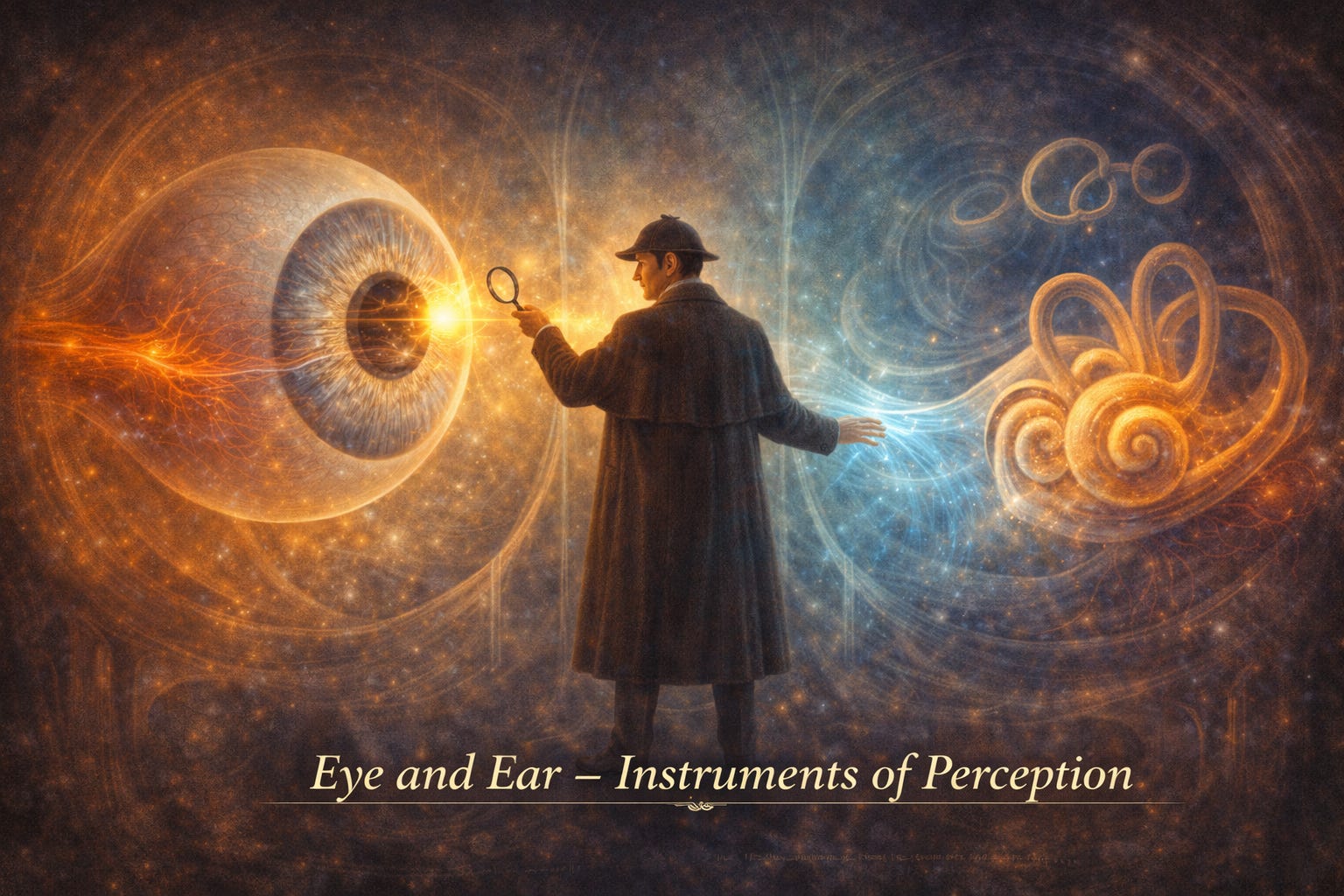 ANAHN 10: Eye and Ear - The Instruments of Perception | If the cranial fossa was a chamber of protection,this episode is a chamber of interpretation.Here, the body performs one of its most extraordinary feats:* It converts light into sight* It transforms vibration into sound* It translates motion into balanceThe eye and ear are not simply organs.They are interfaces between physics and perception.PART I - THE EYE: A LENS THAT THINKSThe Orbit: The Housing of VisionThe orbit is not just a socket - it is a precision-engineered cone:* Seven bones form its walls* It contains:* The eyeball (orb)* Muscles, nerves, vessels, fatThe diagram on page 163 shows this conical structure clearly - walls converging posteriorly, directing all structures toward a common apex.Everything entering the orbit is guided - nothing is random.The Eyeball (Orb): A Layered InstrumentThe eye is built like a three-layered sphere, each with a distinct role:* Fibrous tunic* Sclera (white, protective)* Cornea (transparent window)* Vascular tunic* Choroid (blood supply)* Ciliary body (focus control)* Iris (light regulation)* Retinal tunic* Neural tissue → converts light to signalsThe diagram on page 166 beautifully shows these layers, with the retina lining the inner wall like a sensory screen.The eye is not just a camera - it is a living, adaptive sensor.Light Pathway: The Journey of SightLight passes through a carefully ordered system:* Cornea* Aqueous humour* Lens* Vitreous body* RetinaEach structure refracts light, bending it toward the retina.At the retina:* Rods → detect light intensity* Cones → detect colour and detail* Fovea → highest acuity* Optic disc → blind spotVision is not seen - it is constructed.Accommodation: Focusing the WorldThe lens changes shape via the ciliary muscle:* Contracts → lens becomes convex → near vision* Relaxes → lens flattens → distant visionThis is not conscious - it is autonomic precision.Eye Movements: The Six Directions of ControlSeven extrinsic muscles guide the eye:* 4 recti (up, down, medial, lateral)* 2 obliques (rotational correction)* 1 levator (eyelid)Innervation follows a simple rule:* CN III → most muscles* CN IV → superior oblique* CN VI → lateral rectusLR6 SO4 AO3 - a rule that anchors chaos.Pupillary Control: Light RegulationTwo opposing muscles in the iris:* Sphincter pupillae → constriction (parasympathetic)* Dilator pupillae → dilation (sympathetic)The pupil is not passive - it is a dynamic gatekeeper.Clinical Threads (Eye)* Cataract → lens opacity* Glaucoma → increased intraocular pressure (aqueous humour imbalance)* Retinal detachment → separation from choroid* Myopia/Hyperopia → focusing errorsIn the eye, millimetres define clarity - or blindness.PART II - THE EAR: A SYSTEM OF TRANSLATIONThe Ear’s Three Chambers* External ear → collects sound* Middle ear → amplifies sound* Inner ear → converts sound + detects balanceMiddle Ear: The AmplifierContains three ossicles:* Malleus* Incus* StapesThey:* Transmit vibration from tympanic membrane* Amplify it ~20× before reaching inner earThe diagram on page 176 shows this chain clearly - like a mechanical relay system.Tiny bones, enormous effect.Inner Ear: The Dual System1. Cochlea (Hearing)* Spiral structure (like a snail shell)* Contains organ of Corti* Converts fluid movement → nerve impulses2. Vestibular System (Balance)* Semicircular canals (angular motion)* Utricle & saccule (linear motion)The diagram on page 177 illustrates this beautifully - the cochlea curling forward, canals looping like gyroscopes.Hearing tells you what is happening.Balance tells you where you are.Fluid Dynamics: The Hidden LanguageTwo fluids:* Perilymph (outer)* Endolymph (inner)Movement of these fluids:→ stimulates hair cells→ generates nerve signalsCranial Nerve VIII: The MessengerThe vestibulocochlear nerve carries:* Cochlear division → hearing* Vestibular division → balanceTwo functions, one pathway.Clinical Threads (Ear)* Otitis media → infection via auditory tube* Otosclerosis → stapes fixation* Ménière’s disease → excess endolymph* Neural hearing loss → nerve damageIn the ear, pressure, fluid, and vibration define reality.Key Takeaways* The eye converts light into neural signals via layered structures* The retina is the true sensory surface of vision* Eye movement is coordinated by CN III, IV, and VI* The ear converts vibration into sound and motion into balance* The middle ear amplifies sound; the inner ear transduces it* Fluid movement in the inner ear is central to function* Cranial nerve VIII carries both hearing and balance This is a public episode. If you'd like to discuss this with other subscribers or get access to bonus episodes, visit drmanaankarray.substack.com/subscribe | 53m 13s | ||||||
| 4/30/26 | 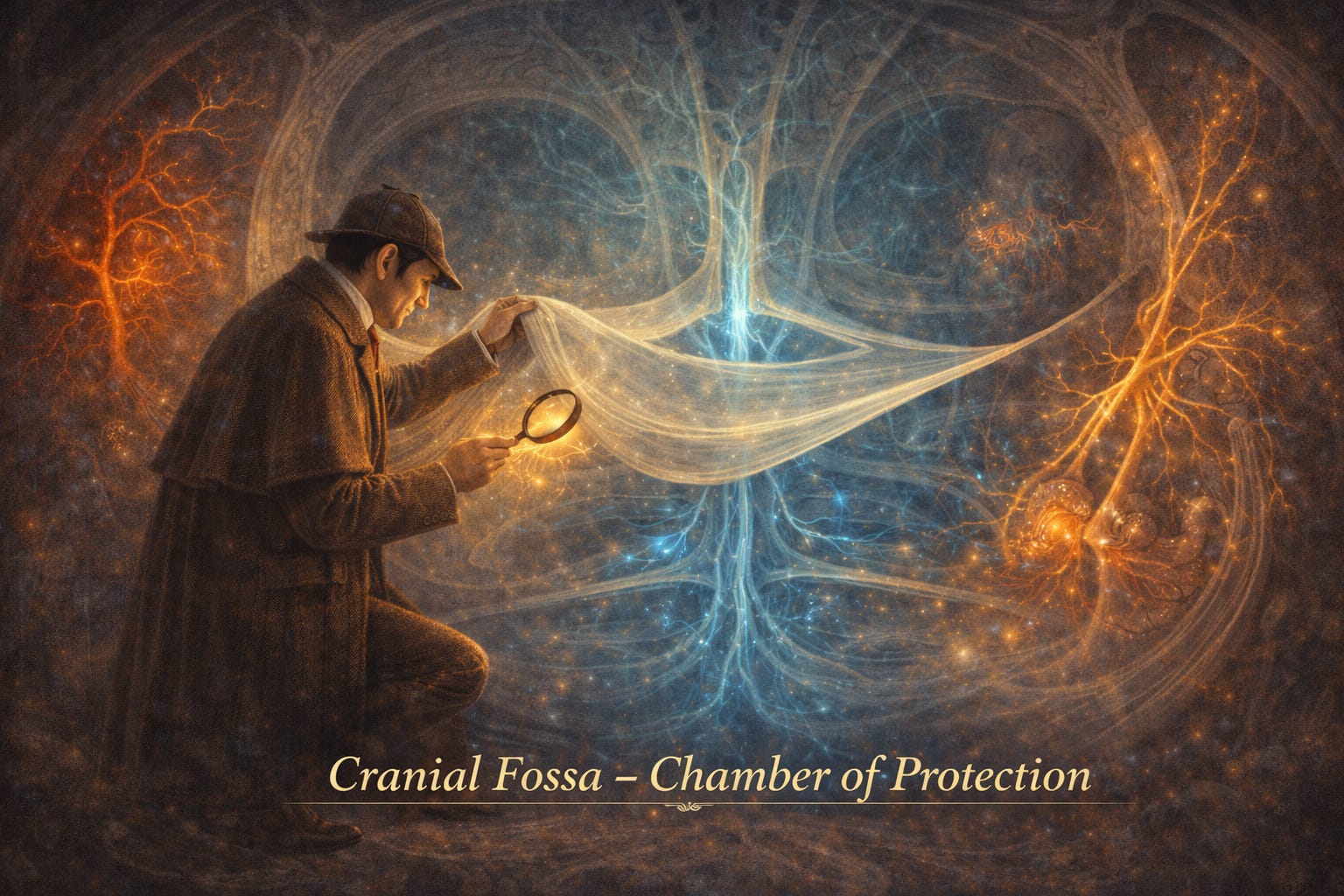 ANAHN 09: Cranial Fossa - The Chamber of Protection and Passage | If the face was a stage, the cranial fossa is the vault beneath it - a protected chamber where the brain rests, suspended within layers of defence, yet threaded with pathways of extraordinary vulnerability.This chapter is not simply about structure.It is about containment, support, and flow.Within the cranial fossa:* The brain is wrapped, not directly by bone, but by layered protection* Blood does not simply circulate - it is channelled through rigid sinuses* Nerves do not wander - they exit through precise gatewaysAnd yet, this chamber is not sealed.It is a space of communication, where extracranial and intracranial worlds meet - with profound clinical implications.The Cranial Fossa: A Protected CavityThe cranial fossa is the internal space of the skull, housing:* The brain* The meninges* The cranial nerves as they emerge and exitIt is not simply a container - it is a structured environment with layers, compartments, and channels.The Meninges: Layers of ProtectionThe brain is enveloped by three layers:* Dura mater (outer, tough layer)* Arachnoid mater* Pia materThis chapter focuses on the dura mater, the most robust protective layerDura Mater: The Dual-Layer ShieldThe dura is not a single sheet - it is composed of two layers:* Periosteal layer (attached to the skull)* Meningeal layer (closely related to the brain surface)These layers:* Adhere tightly at sutures* Separate in specific regions to form venous sinusesThe dura is both armour and architecture.Dural Reflections: Internal PartitionsThe dura folds inward to create reflections - structural supports that stabilise the brain.Key reflections include:* Falx cerebri - separates left and right cerebral hemispheres* Tentorium cerebelli - separates cerebrum from cerebellum* Falx cerebelli - separates cerebellar hemispheres* Diaphragma sella - covers the pituitary glandThese folds:* Provide mechanical support* Create compartments* Form the framework for venous sinusesThe brain is not floating freely - it is gently held within a system of internal scaffolding.Venous Sinuses: Channels Without WallsUnlike normal veins, dural venous sinuses are:* Endothelial-lined spaces (not true vessels)* Rigid and valveless* Formed between layers of duraThey:* Collect blood from the brain, meninges, and skull* Receive cerebrospinal fluid* Drain ultimately into the internal jugular veinKey sinuses include:* Superior sagittal* Inferior sagittal* Straight* Transverse* Sigmoid* CavernousThese are not flexible pipes - they are fixed channels carved into the dura.The Cavernous Sinus: A Critical CrossroadsOne of the most clinically significant spaces:Located beside the sella turcica, it contains:* Internal carotid artery* Abducens nerve (VI)And in its walls:* Oculomotor (III)* Trochlear (IV)* Trigeminal divisions (V1, V2)This is where vessels and nerves travel in intimate proximity - a place where pathology spreads with consequences.Arterial Supply of the DuraThe dura is supplied by meningeal arteries:* Middle meningeal artery (most important)* Anterior meningeal* Accessory meningeal* Posterior meningeal arteriesThe middle meningeal artery:* Enters via the foramen spinosum* Grooves the inner skull* Is a key player in epidural haemorrhageDiploic and Emissary Veins: Hidden ConnectionsDiploic veins:* Located within skull bone* Connect scalp veins, meningeal veins, and sinusesEmissary veins:* Connect extracranial veins with intracranial sinuses* Valveless → bidirectional flowThese veins ignore boundaries - they connect outside and inside worlds.Clinical Insight: Pathways of DangerBecause of valveless systems:* Infection can travel from scalp or face → cranial cavityEpidural haematoma:* Middle meningeal artery rupture* Initial recovery → rapid deterioration* Surgical emergencyCavernous sinus pathology:* Affects multiple cranial nerves* Leads to ophthalmoplegia, sensory lossIn the cranial fossa, pressure is unforgiving - small changes have large consequences.Cranial Nerves: The Exit RoutesThere are 12 cranial nerves, each leaving the cranial cavity via foramina.Examples:* CN I (olfactory) → cribriform plate* CN II (optic) → optic canal* CN V (trigeminal) → divides into V1, V2, V3* CN VII & VIII → internal acoustic meatus* CN IX, X, XI → jugular foramen* CN XII → hypoglossal canalEach nerve is a traveller, leaving the protected chamber to serve the body.Meningeal InnervationThe dura is innervated primarily by:* Trigeminal nerve (CN V)* With contributions from:* Vagus (X)* Hypoglossal (XII)* Upper cervical nervesThis explains:* Why dural irritation causes referred pain (headaches)Key Takeaways* The cranial fossa houses the brain, meninges, and cranial nerve pathways* Dura mater has two layers: periosteal and meningeal* Dural reflections partition and support the brain* Venous sinuses are rigid, valveless channels draining into the internal jugular vein* The cavernous sinus contains critical neurovascular structures* The middle meningeal artery is clinically important in epidural haemorrhage* Emissary veins allow extracranial–intracranial communication* Cranial nerves exit through specific foramina* Dural innervation explains headache patterns This is a public episode. If you'd like to discuss this with other subscribers or get access to bonus episodes, visit drmanaankarray.substack.com/subscribe | 53m 29s | ||||||
| 4/29/26 | 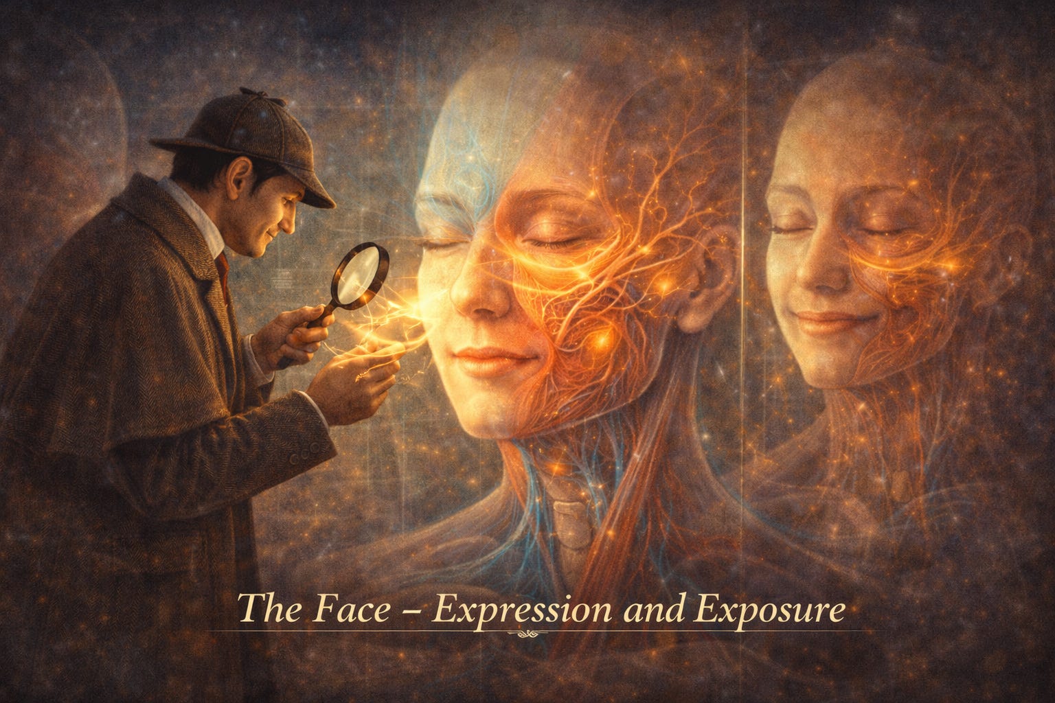 ANAHN 08: The Face - Expression, Emotion, and Exposure | If the neck was a corridor, the face is a stage.But this is not a passive surface.It is a living interface where structure meets meaning.The face is where:* Muscles do not just move - they express* Nerves do not just transmit - they interpret* Blood does not just flow - it reveals life in colour and warmthAnd yet, beneath this expressive surface lies a system that is clinically vulnerable, anatomically intricate, and dangerously connected.The Face: A Functional IdentityThe superficial face is built from:* Thin skin* Highly vascular connective tissue* Interwoven muscles* Dense neural networksUnlike most of the body, the soft tissue is thin over bone, allowing structure to be easily palpated and movement to be highly visibleThe face is not built for protection.It is built for communication.The Defining Feature: Muscles of ExpressionThese muscles are unique in the human body.* They arise from the second pharyngeal arch* They are innervated by the facial nerve (CN VII)* They insert into skin, not boneThis single fact changes everything.When they contract:They do not move joints - they reshape emotion.They gather around orifices:* Eyes* Nose* MouthAnd act in coordinated groups to produce:* Smiles* Frowns* Blinks* Speech articulationThe face is an orchestra where no muscle plays alone.The Scalp: Layers and the “Danger Space”The scalp is not just a covering - it is a layered system:* Skin* Fibroadipose layer* Epicranius muscle (frontalis + occipitalis)* Galea aponeurotica* Loose connective tissue (danger space)* PericraniumThis “danger space” allows movement - but also allows infection to spread deeply.Vascularity: A Face Full of FlowThe face is exceptionally vascular.* Supplied by branches of both:* External carotid artery* Internal carotid artery* Forms extensive anastomotic networksThis leads to two key realities:1. Bleeding is profuse and difficult to control2. Healing is rapid and resilientThe face bleeds easily - but it also recovers beautifully.The Facial Artery: The Signature PathwayThe facial artery takes a tortuous, winding course:* Crosses the mandible* Travels towards the corner of the mouth* Ascends along the nose* Ends near the eye as the angular arteryIts branches supply:* Lips* Nose* CheekIt is a vessel that follows expression itself.Venous System: The Hidden RiskUnlike many veins in the body:* Facial veins are valvelessThis means blood can flow in either direction.And critically:* They connect to the cavernous sinus inside the skullThis is where beauty meets danger.The “Danger Triangle” of the FaceA small region carries disproportionate risk:* Upper lip* Nose* Area to medial eyeInfection here can travel via venous pathways to the cavernous sinus, leading to:* Thrombosis* Brain involvement* Potential deathA seemingly trivial lesion can become a neurological emergency.Sensory Innervation: The Trigeminal MapThe face is exquisitely sensitive due to the trigeminal nerve (CN V).It divides into three territories:* V1 (Ophthalmic): forehead, upper eyelid, nose* V2 (Maxillary): cheek, upper lip* V3 (Mandibular): lower lip, chin, jawThere is overlap, increasing sensitivity and redundancy.The face feels the world in three overlapping maps.Motor Innervation: The Facial Nerve (CN VII)The facial nerve exits the skull and divides into five key branches:* Temporal* Zygomatic* Buccal* Mandibular* CervicalThese branches form a plexus within the parotid gland before radiating outward.This is the nerve of:* Expression* Symmetry* IdentityThe Buccinator: The Hidden WorkerOften overlooked, but essential:* Compresses cheek* Keeps food between teeth* Assists speech and blowingIt is the quiet stabiliser beneath expression.Clinical Insight: When Expression FailsFacial nerve injury (e.g. Bell palsy):* Drooping face* Inability to close eye* Loss of expression* Speech difficultyTrigeminal neuralgia:* Severe facial pain* Triggered by light touch* Involves sensory pathwaysFacial infections:* Can spread intracranially due to venous connectionsThe face is where dysfunction is not hidden - it is seen immediately.Key Takeaways* Facial muscles insert into skin and create expression* All muscles of facial expression are innervated by CN VII* Sensory supply is via the three divisions of CN V* The face is highly vascular with extensive arterial anastomoses* Facial veins are valveless → bidirectional flow* The danger triangle allows infection to spread to the cavernous sinus* The facial nerve branches within the parotid gland* The buccinator is key for mastication and oral control This is a public episode. If you'd like to discuss this with other subscribers or get access to bonus episodes, visit drmanaankarray.substack.com/subscribe | 1h 08m 21s | ||||||
| 4/28/26 | 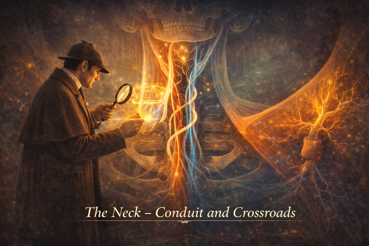 ANAHN 07: The Neck - Conduit, Compass, and Crossroads | If the skull was the fortress, the neck is the gateway system that keeps it alive.This chapter is not about isolated structures - it is about relationships in motion. The neck is a tightly packed corridor where nerves, vessels, muscles, and viscera coexist in remarkable proximity.Everything that sustains the brain must pass through here.Everything that expresses the brain must also emerge from here.The Neck: A Living ConduitThe neck is a cylindrical bridge connecting head to trunk.But more importantly, it is:* A neurovascular highway (brain ↔ body)* A respiratory passage (airflow)* A digestive corridor (swallowing pathway)It is not designed for space.It is designed for efficiency under constraint.Layers: From Surface to DepthThis region is best understood like a careful dissection - layer by layer.Superficial layer:* Skin, platysma* Superficial veins* Cutaneous nervesDeep fascia (the organisers):* Investing fascia → wraps entire neck* Pretracheal fascia → surrounds viscera* Prevertebral fascia → stabilises spine and deep muscles* Carotid sheath → encloses vital structuresThe fascia are not passive coverings - they are compartments of meaning, separating and protecting function.The Great Divider: SternocleidomastoidThe sternocleidomastoid (SCM) is not just a muscle - it is a mapmaker.It divides the neck into two major regions:* Anterior triangle* Posterior triangleThis single muscle transforms complexity into something navigable.Posterior Triangle: The Nerve FieldThis region is a neural landscape.Key contents:* Accessory nerve (CN XI) → motor to SCM & trapezius* Cervical plexus → sensory to neck* Brachial plexus (roots emerging) → upper limb supply* Subclavian artery branchesThe accessory nerve here is vulnerable - injury leads to shoulder droop and impaired head rotation.This triangle teaches a principle:Where nerves travel superficially, function becomes fragile.Anterior Triangle: The Vital CorridorIf the posterior triangle is neural, the anterior triangle is visceral and vascular.Key structures:* Carotid arteries → blood to brain* Internal jugular vein → venous drainage* Thyroid and parathyroid glands* Vagus nerve (CN X)* Cervical sympathetic chainThis is where survival flows - literally.The Carotid Sheath: A Bundle of LifeRunning deep within the neck is a vertical column containing:* Common/Internal carotid artery* Internal jugular vein* Vagus nerveThink of this as a three-strand lifeline:Flow in. Flow out. Regulation between.Muscles: Movement and StabilityThe neck balances mobility and protection.Key groups:* Sternocleidomastoid → rotates and flexes head* Trapezius → supports shoulder and posture* Scalenes → assist breathing, elevate ribs* Infrahyoid muscles → stabilise hyoid and larynx* Prevertebral muscles → deep stabilisersThese muscles do not just move the head - they maintain alignment between intention and action.Arteries: The Ascending SupplyBlood supply ascends like a branching tree.* Common carotid artery → divides into:* Internal carotid (brain)* External carotid (face and neck)Branches include:* Facial artery* Lingual artery* Occipital artery* Maxillary arteryMeanwhile:* Subclavian artery feeds deeper and posterior structuresVeins: The Returning FlowThe internal jugular vein is the main drainage pathway:* Begins at skull base* Drains brain, face, neck* Empties into brachiocephalic veinArteries bring urgency.Veins restore balance.Nerves: The Command NetworkThe neck hosts an intricate neural system:* Cranial nerves (e.g. vagus, accessory)* Cervical plexus (C1–C4)* Brachial plexus (C5–T1 emerging)* Ansa cervicalis → motor to infrahyoid musclesThese networks coordinate:* Movement* Sensation* Autonomic controlThe Cervical Triangles: Maps Within MapsEach triangle contains predictable anatomy.* Posterior triangle → nerves and arteries* Anterior triangle → vessels, glands, visceraThese triangles are not just regions - they are clinical navigation tools.Clinical Insight: Why This MattersThe neck is a high-risk, high-value region:* Surgical procedures (e.g. thyroid surgery) require precision* Trauma can affect airway, vessels, or nerves simultaneously* Fascial spaces allow infections to spread rapidlyUnderstanding anatomy here is not optional - it is protective knowledge.Key Takeaways* The neck is a conduit for neurovascular, respiratory, and digestive systems* Deep fascia organise the region into functional compartments* Sternocleidomastoid divides the neck into anterior and posterior triangles* Posterior triangle is nerve-rich; anterior triangle is vessel- and organ-rich* Carotid sheath contains artery, vein, and vagus nerve* Arterial supply arises from carotid and subclavian systems* Internal jugular vein is the main venous drainage* Cervical and brachial plexuses coordinate sensation and movement* Cervical triangles provide essential clinical landmarks This is a public episode. If you'd like to discuss this with other subscribers or get access to bonus episodes, visit drmanaankarray.substack.com/subscribe | 55m 43s | ||||||
| 4/27/26 |  ANAHN 06: Osteology of the Head and Neck | If embryology was the negotiation, osteology is the contract - fixed, structured, and enduring.This chapter shifts us from possibility to precision. The soft choreography of development has now hardened into bone, and every ridge, foramen, and articulation carries a purpose.The Skull: A Fortress with One DoorThe skull is composed of 22 bones, divided into:* Cranium (8 bones) → protects the brain* Face (14 bones) → shapes identity and functionAlmost all are fused by sutures - immovable joints.Except one.The mandible stands alone - the only movable bone, turning structure into function.Seeing the Skull: Perspectives as UnderstandingThe skull must be studied from multiple views - because no single angle tells the full story.From the anterior view, you see:* Orbits* Nasal aperture* Dental archesFrom the lateral view, you appreciate:* Sutures (coronal, sagittal, lambdoid)* Temporal and infratemporal fossae* Zygomatic archFrom the base, you discover something deeper:The skull is not just a shell - it is a gateway system.The Foramina: Doors in the FortressThe base of the skull is perforated by numerous openings - each a carefully placed gateway for nerves and vessels.For example:* Foramen magnum → brainstem, vertebral arteries* Jugular foramen → CN IX, X, XI* Optic canal → optic nerve* Foramen ovale → mandibular division of trigeminal nerveThese are not random holes - they are organised exits and entries, each with clinical significance.The Orbit: A Seven-Bone ChamberThe orbit is formed by seven bones, creating a protective cavity for the eye .It is shaped like a truncated pyramid:* Wide anteriorly* Narrow posteriorlyKey passageways:* Superior orbital fissure → CN III, IV, VI, V1* Inferior orbital fissure → V2 and vesselsThis is less a cavity and more a neurovascular crossroads.The Nasal Cavity: Structured AirflowPositioned centrally, the nasal cavity is divided by the septum and shaped by:* Ethmoid (superior/medial)* Maxilla and palatine bones (floor)The lateral walls contain:* Conchae (turbinates)* Meatuses (air passages)These structures transform airflow into something purposeful - warming, filtering, directing.The Mandible: Movement in a Static WorldThe mandible is:* Horseshoe-shaped* The only mobile bone of the skull* Articulates at the temporomandibular joint (TMJ)It enables:* Mastication* Speech* ExpressionLook at the mental foramen (see labelled diagram on page 95): it transmits the mental nerve and vessels - an essential landmark in dentistry and surgery .This bone is where structure meets action.The Hyoid: The Floating AnchorThe hyoid bone is unique:* U-shaped* Does not articulate with any other bone* Suspended by muscles and ligamentsIt acts as a functional anchor for:* Tongue* Larynx* Swallowing mechanismsIt is not fixed - yet it is essential.Cervical Vertebrae: Mobility with ControlThe neck introduces controlled movement.There are 7 cervical vertebrae:* C1 (Atlas) → supports the skull* C2 (Axis) → provides rotation via the densTogether, they create:* Flexion/extension (nodding)* Rotation (shaking head “no”)The atlas has no body - just an arch.The axis has a tooth-like projection - the dens.Movement emerges not from strength - but from clever design.Internal Base of the Skull: The Three Tiers of the BrainThe internal skull base forms three cranial fossae:* Anterior cranial fossa → frontal lobes* Middle cranial fossa → temporal lobes, sella turcica (pituitary)* Posterior cranial fossa → brainstem, cerebellumEach is lower than the one before - like descending terraces.This is not just support - it is organisation of the brain itself.Radiological PerspectiveThe radiographs (see pages 96–100) translate anatomy into clinical vision:* Fractures* Sinus opacification* Alignment of cervical spineAnatomy is not complete until it can be seen in shadow and density.Key Takeaways* The skull is composed of 22 bones - mostly fused, with the mandible as the only movable element* The skull must be understood in multiple views to appreciate its complexity* Foramina are structured gateways for neurovascular structures* The orbit is a complex seven-bone cavity with critical fissures* The nasal cavity is designed for airflow conditioning* The mandible enables function - chewing, speech, movement* The hyoid is a suspended anchor for swallowing and speech* The atlas and axis enable controlled head movement* The cranial fossae organise the brain into functional compartments This is a public episode. If you'd like to discuss this with other subscribers or get access to bonus episodes, visit drmanaankarray.substack.com/subscribe | 41m 59s | ||||||
| 4/26/26 |  ANAHN 05: Embryology of the Head and Neck | If Episode 4 showed us the finished cathedral, Chapter 5 reveals the construction site - and it is anything but orderly.The head and neck do not simply “grow.” They assemble, fuse, migrate, and transform - often within narrow windows of time where a single misstep can echo for life.At the centre of this process lies a deceptively simple idea:Development is not just about what forms - it is about when, where, and with what signals it forms.The Language of Development: A Borrowed VocabularyEmbryology uses terms like branchial (gill-related), reflecting our evolutionary inheritance. Humans do not form gills, but the pharyngeal system is their structural descendant .This matters because it reframes anatomy not as isolated parts - but as adapted remnants of older blueprints.Genetic Orchestration: The Timing CodeDevelopment is governed by:* Homeobox (Hox) genes → control spatial patterning* Signalling molecules → guide cell behaviour* Induction → one group of cells directing anotherThese create a “window of opportunity” - a precise developmental moment when an event must occur .Miss that window - and the structure may never form correctly.Think of this as a symphony without a conductor - only timing rules. If one instrument enters too early or too late, the entire piece changes.The Pharyngeal Apparatus: The Foundational BlueprintAround weeks 4–5, the embryo develops a repeating system:* Pharyngeal arches (mesenchyme)* Pouches (endoderm)* Grooves (ectoderm)Each arch is not just a structure - it is a functional unit, containing:* Bone/cartilage* Muscle* Artery* Cranial nerveThis is one of the most important organising principles in head & neck anatomy.The Pharyngeal Arches: A Pattern That PersistsThere are five functional arches (I, II, III, IV, VI - V disappears). Each gives rise to predictable derivatives:Arch I (Mandibular) – CN V* Jaw, maxilla, malleus, incus* Muscles of masticationArch II (Hyoid) – CN VII* Facial expression muscles* Stapes, styloid processArch III – CN IX* Stylopharyngeus* Part of hyoidArch IV & VI – CN X* Laryngeal cartilages* Pharyngeal and laryngeal musclesThis pattern explains something profound:Adult anatomy is a memory of embryological segmentation.Grooves and Pouches: Hidden Contributors* 1st groove → external auditory meatus* 1st pouch → middle ear cavity + auditory tube* 2nd pouch → palatine tonsil* 3rd pouch → thymus + inferior parathyroids* 4th pouch → superior parathyroids* 5th pouch → thyroid parafollicular cellsThese are not obvious when looking at an adult - but they explain many clinical anomalies.The Tongue: A Composite StructureThe tongue forms from multiple arches:* Anterior 2/3 → Arch I* Posterior 1/3 → Arches II–IV* Muscles → migrate from occipital somites (CN XII)This explains its complex innervation - a layered story of origins rather than a single design.The Thyroid: A Migratory StoryThe thyroid begins at the foramen cecum and descends into the neck via the thyroglossal duct .If this migration is incomplete:* Thyroglossal cysts* Lingual thyroidDevelopment is not just formation - it is movement with memory.Face Formation: Fusion as IdentityThe face emerges from five key processes:* Frontonasal prominence* Paired maxillary processes* Paired mandibular processesFusion is everything:* Median nasal processes → intermaxillary segment* Forms philtrum, primary palate, central upper lipFailure of fusion leads to visible clinical conditions.The Palate: Separation of Two WorldsInitially, oral and nasal cavities are one.Then:* Palatal shelves grow downward* Elevate above the tongue* Fuse in the midline → secondary palateThis is a critical developmental moment - a hinge point between normal function and pathology.Clinical Insight: When Timing FailsCleft LipFailure of fusion between maxillary process and intermaxillary segmentCleft PalateFailure of palatal shelves to fuseCervical cystsPersistence of pharyngeal groovesTreacher Collins syndromeFirst arch developmental failureThese are not random defects - they are missed conversations in development.Key Takeaways* Development is governed by timing (windows), signalling, and gene activation* Pharyngeal arches are the fundamental organisational units* Each arch has its own nerve, muscle, and skeletal derivatives* Pouches and grooves contribute to hidden but clinically vital structures* The tongue and thyroid reflect multi-origin and migratory development* The face forms through fusion of multiple embryonic processes* The palate separates oral and nasal cavities - failure leads to clefting* Many clinical conditions are failures of timing, fusion, or migration This is a public episode. If you'd like to discuss this with other subscribers or get access to bonus episodes, visit drmanaankarray.substack.com/subscribe | 58m 53s | ||||||
| 4/25/26 |  ANAHN 04: The Oral Cavity, Palate, and Pharynx | This episode brings us to one of the most clinically alive regions of anatomy - the oral cavity and its continuation into the pharynx. This is not just a space; it is a gateway. Everything that sustains life - air, food, communication - passes through here.At its simplest, the oral cavity is divided into two spaces:* The vestibule (between lips/cheeks and teeth)* The oral cavity proper (within the dental arches)This distinction is subtle but powerful. It explains how dentists examine, how infections spread, and how local anaesthesia is targeted.The Lips: The ThresholdThe journey begins at the lips - highly vascular, mobile structures guarding the entrance. Their red vermilion zone is not just aesthetic; it reflects a rich blood supply beneath thin epithelium .Clinically, they are a fusion point of embryological processes - explaining conditions like cleft lip.Think of the lips as the gatekeepers - flexible, expressive, and protective.The Vestibule: The Outer ChamberThe vestibule is the space between lips/cheeks and teeth. It is not empty - it is active:* Receives saliva (e.g., parotid duct opening near the molars)* Contains minor salivary glands* Houses structures like frenula and mucosal foldsThis is where injections are placed, where swelling is first seen, and where anatomy is palpated.The Oral Cavity Proper: The Functional CoreInside the dental arches lies the true working chamber:* Roof: palate* Floor: tongue* Posterior boundary: oropharyngeal isthmus (fauces)This is where chewing, speech shaping, and early swallowing begin.The Tongue: The Architect of FunctionThe tongue is not just a muscle - it is a multifunctional organ divided into:* Anterior 2/3 (body) – oral cavity* Posterior 1/3 (base) – pharynxThe dividing line - the sulcus terminalis - is more than anatomical; it marks a shift in function, innervation, and embryology.Its surface is specialised:* Filiform papillae → texture (no taste)* Fungiform & circumvallate → taste* Lingual tonsils posteriorly → immune roleThe tongue is where movement meets sensation meets immunity.The Palate: The Divider of WorldsThe palate forms the roof of the oral cavity and is divided into:* Hard palate (anterior, bony)* Soft palate (posterior, muscular)Its role is profound: it separates the oral and nasal cavities, allowing breathing and eating to coexist without chaos.During swallowing, the soft palate elevates - sealing the nasopharynx.A simple act, but one that prevents aspiration and enables coordinated swallowing.Teeth: The Functional BoundaryTeeth are not just for chewing - they define spaces:* Separate vestibule from oral cavity proper* Provide mechanical breakdown of food* Anchor within alveolar bone via periodontal ligamentTwo dentitions exist:* Deciduous (20 teeth)* Permanent (32 teeth)Their development (odontogenesis) follows a staged process - bud, cap, bell - reflecting increasing complexity and differentiation.The Pharynx: The CrossroadsBeyond the oral cavity lies the pharynx - a shared corridor for air and food.It extends from the base of the skull to the oesophagus and is divided into:* Nasopharynx (air only)* Oropharynx (air + food)* Laryngopharynx (towards oesophagus)Its muscular walls (pharyngeal constrictors) act in sequence - guiding food downward.Here lies a crucial idea:The pharynx is not just a tube - it is a decision point, directing life-sustaining pathways.Clinical Insight: Why This Chapter MattersThis region is where multiple systems converge:* Airway obstruction* Swallowing disorders* Speech production* Infection spread (tonsils, lymphatic ring of Waldeyer)* Dental and surgical accessEven a simple action like saying “ah” lifts the palate and reveals the oropharynx - a reminder that anatomy is always in motion.Key Takeaways* Oral cavity = vestibule + oral cavity proper* Lips form the vascular, protective entrance* Vestibule is a clinically important space for examination and procedures* Oral cavity proper is bounded by palate (roof) and tongue (floor)* Tongue is divided into anterior (oral) and posterior (pharyngeal) parts* Palate separates oral and nasal cavities; soft palate enables swallowing* Teeth define functional boundaries and develop through staged embryology* Pharynx is a shared airway and пищ pathway divided into three regions* Coordination between these structures enables speech, swallowing, and breathing This is a public episode. If you'd like to discuss this with other subscribers or get access to bonus episodes, visit drmanaankarray.substack.com/subscribe | 1h 06m 51s | ||||||
| 4/24/26 |  ANAHN 03: Body Systems | This chapter steps back from regions and maps the body as a living network of systems - each one specialised, yet deeply interdependent. If Chapter 2 gave us the language of anatomy, this chapter gives us its living context.We begin at the smallest scale: the cell, the fundamental unit of life. Cells organise into tissues, tissues into organs, and organs into systems. This hierarchy is not merely structural - it is functional. Each level represents increasing coordination, a quiet orchestration of purpose.From here, the chapter surveys the major systems that shape the head and neck.The integumentary system forms the body’s boundary - protective, sensory, and regulatory. It is not just a covering, but an interface between the internal world and the external environment. Within it, layers emerge: epidermis, dermis, and hypodermis - each contributing to protection, sensation, and adaptation.The muscular system introduces movement. Here, three distinct types - skeletal, cardiac, and smooth - demonstrate how form dictates function. In the head and neck, muscles take on added nuance: some move bone, others move expression itself. The idea of origin, insertion, and coordinated action begins to take shape.The skeletal system provides structure and protection, but also serves as a dynamic organ - storing minerals, producing blood cells, and adapting continuously to stress. Bone is not static; it remodels in response to the forces placed upon it.The circulatory system brings flow - transporting oxygen, nutrients, and waste. It is both a delivery network and a communication system, linking distant regions into a unified whole. Alongside it, the lymphatic system filters and defends, quietly maintaining internal balance.Finally, the nervous system emerges as the master integrator. It perceives, processes, and responds. Divided into central and peripheral components, and further into voluntary and autonomic control, it governs both conscious action and unconscious regulation. Within this, the balance between sympathetic (action) and parasympathetic (restoration) systems reflects a deeper principle: stability through opposition.This chapter is not about memorising systems in isolation. It is about recognising that every structure in the head and neck exists within these systems - receiving blood, responding to nerves, supported by bone, moved by muscle, and protected by skin.Understanding systems transforms anatomy from a static map into a living, dynamic network.Key Takeaways* The body is organised hierarchically: cells → tissues → organs → systems* Structure and function are inseparable at every level of organisation* The integumentary system protects, senses, and regulates the body* The muscular system enables movement through coordinated contraction:* Skeletal (voluntary)* Cardiac (rhythmic, involuntary)* Smooth (visceral, involuntary)* The skeletal system provides support, protection, leverage, mineral storage, and blood formation* Bone is dynamic and remodels in response to stress* The circulatory system transports oxygen, nutrients, hormones, and waste* The lymphatic system filters fluid and contributes to immune defence* The nervous system integrates and controls body function:* CNS (brain and spinal cord)* PNS (cranial and spinal nerves)* The autonomic system balances:* Sympathetic (“fight or flight”)* Parasympathetic (“rest and restore”)* All head and neck structures are expressions of these interacting systems This is a public episode. If you'd like to discuss this with other subscribers or get access to bonus episodes, visit drmanaankarray.substack.com/subscribe | 1h 20m 34s | ||||||
| 4/23/26 |  ANAHN 02: Anatomic Concepts | This chapter is the grammar of anatomy - the quiet framework that allows everything else to make sense. Before we explore structures, we must first understand how anatomists think, describe, and orient themselves within the human body.At its core, anatomy is a spatial science. It does not simply name structures; it describes their relationships. To do this effectively, a universal reference point is required - the anatomic position. From this standardised stance, every direction, movement, and relationship becomes meaningful and consistent.The chapter then introduces the divisions of anatomy, reminding us that the body can be studied at different scales and through different lenses: from the microscopic world of tissues to the visible architecture of gross anatomy, and from developmental origins to the intricate wiring of the nervous system. These are not separate disciplines, but different windows into the same structure.A crucial distinction emerges between systemic and regional anatomy. While systemic anatomy isolates systems for clarity, regional anatomy restores the body to its natural complexity. Nowhere is this more important than in the head and neck, where structures are densely packed and deeply interconnected.The language of anatomy is then built through descriptive terms - anterior and posterior, medial and lateral, proximal and distal. These are not just labels; they are coordinates in a three-dimensional map. Alongside these terms come the planes of the body - sagittal, coronal, and transverse - which allow us to slice the body conceptually and understand it layer by layer.Finally, the chapter introduces a humbling but essential truth: variation is normal. The human body does not always follow the textbook. Subtle differences in vessels, nerves, and structures are common, and true anatomical understanding lies not in memorising a single pattern, but in recognising and interpreting variation.This chapter teaches you how to see, how to orient, and how to describe. Without it, anatomy is a list. With it, anatomy becomes a map.Key Takeaways* Anatomy is a spatial language that describes relationships between structures* The anatomic position is the universal reference point for all descriptions* Anatomy is divided into:* Developmental (formation of the body)* Neuroanatomy (nervous system)* Microscopic (histology)* Macroscopic (gross anatomy)* Gross anatomy can be studied in two ways:* Systemic (by systems)* Regional (by body areas)* The head and neck are best understood using a regional approach due to structural complexity* Descriptive terms (anterior, posterior, medial, lateral, proximal, distal, superficial, deep) form the coordinate system of anatomy* The body is understood through three key planes:* Sagittal (left/right division)* Coronal (front/back division)* Transverse (upper/lower division)* Anatomic variation is common and must be recognised and interpreted clinically This is a public episode. If you'd like to discuss this with other subscribers or get access to bonus episodes, visit drmanaankarray.substack.com/subscribe | 40m 18s | ||||||
| 4/22/26 |  ANAHN 01: Introduction to Head and Neck Anatomy | This opening chapter is not about structures - it is about seeing. It traces the long arc of anatomical curiosity, from early cranial surgery thousands of years ago to the disciplined dissections of Renaissance Europe. What begins as fascination becomes method, and what begins as observation becomes science.We follow the evolution of anatomical thought: from early Greek philosophers who linked structure to function, through the persistence of humoral theory, to the revolutionary clarity brought by figures like Vesalius and Harvey. The chapter reveals that anatomy is not static knowledge - it is a story of correction, refinement, and deeper understanding.Crucially, this chapter introduces the ways of organising anatomy: systemic, regional, and surgical. While systemic anatomy separates the body into neat divisions, the head and neck resist such simplification. Their structures are tightly interwoven - nerves, vessels, muscles, and spaces layered in close proximity.This is why the book - and this entire series - adopts a regional approach. Instead of isolating systems, it teaches anatomy as it exists in reality: interconnected, spatially complex, and clinically meaningful.This chapter therefore does something subtle but powerful: it shifts the learner from memorising parts to thinking in relationships. It prepares you not just to learn anatomy, but to navigate it. Key Takeaways* Anatomy has evolved over millennia - from early surgical practices to modern scientific discipline* Early thinkers (e.g., Alcmaeon, Aristotle) began linking structure to function, forming the foundation of modern anatomy* Historical misconceptions (e.g., humoral theory) remind us that medical knowledge is constantly refined* The Renaissance marked a turning point with systematic dissection and accurate anatomical illustration* There are three main approaches to anatomy:* Systemic (organised by systems)* Regional (organised by body areas)* Surgical (clinically applied regional anatomy)* The head and neck demand a regional approach due to dense structural interrelationships* True understanding comes from seeing relationships, not isolated structures This is a public episode. If you'd like to discuss this with other subscribers or get access to bonus episodes, visit drmanaankarray.substack.com/subscribe | 32m 33s | ||||||
| 4/21/26 |  GPH 109: The Future of International Public Health | International public health is entering a period of profound transformation. Globalisation, climate change, demographic shifts, pandemics, digital surveillance technologies, and geopolitical realignments are reshaping both risk and response.This chapter examines the evolving role of multilateral organisations, global financing mechanisms, pandemic preparedness frameworks, and cross-border governance. It explores innovation in data systems, vaccine development platforms, global surveillance networks, and health diplomacy.The future of international public health depends not only on technical expertise but on political will, equitable resource distribution, and global solidarity. Preparedness must move from reactive crisis response to sustained structural resilience.Global health security and global health equity must advance together.Key Takeaways* Global health faces emerging risks including climate change and pandemics.* International governance structures continue to evolve.* Data systems and surveillance technologies are transforming response capacity.* Health diplomacy plays a critical role in cooperation.* Preparedness requires long-term investment.* Equity must remain central to global health strategy.* Resilience depends on coordination and sustained political commitment. This is a public episode. If you'd like to discuss this with other subscribers or get access to bonus episodes, visit drmanaankarray.substack.com/subscribe | 1h 11m 36s | ||||||
| 4/20/26 |  GPH 108: Private Support of Public Health | Public health is not funded or delivered by governments alone. Philanthropic foundations, private donors, non-governmental organisations, corporate partners, and social enterprises play an increasingly prominent role in financing, research, service delivery, and advocacy.This chapter examines the evolution of private sector engagement in public health, including funding mechanisms, public–private partnerships, global health initiatives, and the role of foundations in shaping priorities. It also addresses governance, transparency, accountability, and potential conflicts of interest.Private contributions can accelerate innovation and mobilise resources at scale - but they must align with population needs, equity principles, and national strategies.Public health thrives when partnership is principled.Key Takeaways* Private actors contribute funding, innovation, and service capacity.* Public–private partnerships can strengthen system delivery.* Philanthropic funding influences global health priorities.* Governance and accountability mechanisms are essential.* Conflicts of interest must be managed transparently.* Alignment with equity and national strategy is critical.* Partnership requires clarity of roles and shared goals. This is a public episode. If you'd like to discuss this with other subscribers or get access to bonus episodes, visit drmanaankarray.substack.com/subscribe | 56m 32s | ||||||
| 4/20/26 |  GPH 107: Environmental/ Chemical/ Radiation Emergency Response | Environmental, chemical, and radiation emergencies present complex and potentially invisible threats to population health. Industrial spills, toxic releases, nuclear incidents, and accidental exposures demand rapid assessment, specialised expertise, and coordinated containment.This chapter explores hazard identification, exposure pathways, toxicology, dose assessment, decontamination protocols, evacuation planning, risk communication, and long-term monitoring. It examines inter-agency coordination, regulatory oversight, and preparedness planning.Unlike infectious outbreaks, these threats may be silent and odourless, requiring measurement instruments rather than clinical suspicion alone. Effective response depends on preparedness infrastructure, clear communication, and evidence-based thresholds for action.In environmental emergencies, precision protects.Key Takeaways* Environmental emergencies require rapid hazard identification and containment.* Exposure assessment guides protective action.* Decontamination and evacuation protocols reduce risk.* Toxicology and dose–response principles inform response decisions.* Inter-agency coordination is essential.* Risk communication must be clear and transparent.* Long-term monitoring ensures sustained protection. This is a public episode. If you'd like to discuss this with other subscribers or get access to bonus episodes, visit drmanaankarray.substack.com/subscribe | 1h 09m 15s | ||||||
| 4/18/26 |  GPH 106: Emergency Public Health and Humanitarian Assistance | Emergencies - whether natural disasters, armed conflict, epidemics, or sudden displacement - disrupt infrastructure, overwhelm health systems, and expose populations to acute risk. Public health in emergencies requires speed, coordination, and ethical clarity.This chapter explores rapid health needs assessment, emergency surveillance, outbreak control, water and sanitation provision, food security, shelter, vaccination campaigns, and coordination across agencies. It examines humanitarian principles, cluster coordination models, and the interface between national authorities and international responders.Emergency public health is not improvisation; it is organised readiness. Prepared systems, trained personnel, and clear governance structures determine whether crises escalate or stabilise.Response capacity is a measure of system strength.Key Takeaways* Emergencies disrupt infrastructure and increase health vulnerability.* Rapid health needs assessment guides prioritisation.* Surveillance and outbreak control are critical in crisis settings.* Water, sanitation, shelter, and nutrition are core public health functions in emergencies.* Coordination across agencies improves efficiency and equity.* Humanitarian principles guide ethical response.* Preparedness determines resilience. This is a public episode. If you'd like to discuss this with other subscribers or get access to bonus episodes, visit drmanaankarray.substack.com/subscribe | 56m 46s | ||||||
Showing 25 of 562
Sponsor Intelligence
Sign in to see which brands sponsor this podcast, their ad offers, and promo codes.
Chart Positions
5 placements across 4 markets.
Chart Positions
5 placements across 4 markets.