
Insights from recent episode analysis
Audience Interest
Podcast Focus
Publishing Consistency
Platform Reach
Insights are generated by CastFox AI using publicly available data, episode content, and proprietary models.
Most discussed topics
Brands & references
Total monthly reach
Estimated from 30 chart positions in 30 markets.
By chart position
- 🇬🇧GB · Medicine#22100K to 300K
- 🇦🇺AU · Medicine#33100K to 300K
- 🇨🇦CA · Medicine#7630K to 100K
- 🇺🇸US · Medicine#7730K to 100K
- 🇮🇳IN · Medicine#7100K to 300K
- Per-Episode Audience
Est. listeners per new episode within ~30 days
285K to 883K🎙 ~2x weekly·228 episodes·Last published 2w ago - Monthly Reach
Unique listeners across all episodes (30 days)
569K to 1.8M🇬🇧17%🇦🇺17%🇮🇳17%+27 more - Active Followers
Loyal subscribers who consistently listen
228K to 706K
Market Insights
Platform Distribution
Reach across major podcast platforms, updated hourly
Total Followers
—
Total Plays
—
Total Reviews
—
* Data sourced directly from platform APIs and aggregated hourly across all major podcast directories.
On the show
From 12 epsHosts
Recent guests
No guests detected in recent episodes.
Recent episodes
Episode 224: Kidney Stones
Jun 8, 2026
Episode 223: Thyroid Storm
May 15, 2026
9m 16s
Episode 222: Local Anesthetic Systemic Toxicity (LAST)
Apr 7, 2026
Episode 221: High-Output Heart Failure
Mar 24, 2026
Episode 220: Post-ROSC Care
Mar 3, 2026
Social Links & Contact
Official channels & resources
Official Website
Login
RSS Feed
Login
| Date | Episode | Topics | Guests | Brands | Places | Keywords | Sponsor | Length | |
|---|---|---|---|---|---|---|---|---|---|
| 6/8/26 |  Episode 224: Kidney Stones✨ | kidney stonesrenal colic+4 | — | Core EM | — | kidney stonesrenal colic+5 | — | — | |
| 5/15/26 |  Episode 223: Thyroid Storm✨ | Thyroid StormDiagnosis+3 | — | Thyroid Storm | — | Thyroid StormDiagnosis+3 | — | 9m 16s | |
| 4/7/26 | 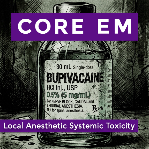 Episode 222: Local Anesthetic Systemic Toxicity (LAST)✨ | local anesthesiasystemic toxicity+3 | — | MidazolamBradycar+1 | — | local anestheticsystemic toxicity+3 | — | — | |
| 3/24/26 |  Episode 221: High-Output Heart Failure✨ | healthmedicine+3 | — | Core EM Modular CME CourseNYU+1 | — | diagnosistreatment+3 | — | — | |
| 3/3/26 |  Episode 220: Post-ROSC Care✨ | Post-ROSC careEmergency medicine+3 | — | Core EM Modular CME CourseNYU+2 | Denmark | ROSCcare optimization+3 | — | — | |
| 2/3/26 |  Episode 219: Meningitis 2.0✨ | bacterial meningitisemergency medicine+3 | — | Core EM Modular CME CourseRifampin+3 | — | bacterial meningitisemergency department+3 | — | — | |
| 1/17/26 |  Episode 218: Sympathetic Crashing Acute Pulmonary Edema (SCAPE)✨ | pulmonary edemaemergency medicine+3 | — | Core EM Modular CME CourseDuonebs+5 | — | SCAPEdiagnosis+3 | — | 12m 45s | |
| 1/1/26 |  Episode 217: Prehospital Blood Transfusion✨ | prehospital careblood transfusion+3 | — | Core EM Modular CME CourseCore Emergency Medicine+8 | USLA County+7 | prehospital bloodshock treatment+3 | — | — | |
| 12/1/25 |  Episode 216: BRUE (Brief Resolved Unexplained Event)✨ | healthmedicine+1 | — | — | — | BRUEBrief Resolved Unexplained Events+3 | — | — | |
| 11/1/25 |  Episode 215: Marburg Virus and Global EM✨ | Marburg VirusGlobal Emergency Medicine+3 | — | WHOAfrica CDC+2 | RwandaUS | Marburg Virus OutbreakRwanda+3 | — | — | |
Want analysis for the episodes below?Free for Pro Submit a request, we'll have your selected episodes analyzed within an hour. Free, at no cost to you, for Pro users. | |||||||||
| 10/2/25 |  Episode 214: Acute Pulmonary Embolism✨ | emergency medicinepulmonary embolism+3 | — | Alteplase | USA | acute pulmonary embolismED management+3 | — | — | |
| 9/1/25 |  Episode 213: Pneumothorax✨ | pneumothoraxrisks+3 | — | — | — | pneumothoraxrisks+3 | — | — | |
| 8/2/25 |  Episode 212: Angioedema | Angioedema – Recognition and Management in the ED Hosts: Maria Mulligan-Buckmiller, MD Brian Gilberti, MD https://media.blubrry.com/coreem/content.blubrry.com/coreem/Angioedema.mp3 Download Leave a Comment Tags: Airway Show Notes Definition & Pathophysiology Angioedema = localized swelling of mucous membranes and subcutaneous tissues due to increased vascular permeability. Triggers increased vascular permeability → fluid shifts into tissues. Etiologies Histamine-mediated (anaphylaxis) Associated with urticaria/hives, pruritus, and redness. Triggered by allergens (foods, insect stings, medications). Rapid onset (minutes to hours). Bradykinin-mediated Hereditary angioedema (HAE): C1 esterase inhibitor deficiency (autosomal dominant). Acquired angioedema: Associated with B-cell lymphoma, autoimmune disease, MGUS. Medication-induced: Most commonly ACE inhibitors; rarely ARBs. Typically lacks urticaria and itching. Gradual onset, can last days if untreated. Idiopathic angioedema Unknown cause; diagnosis of exclusion. Clinical Presentations Swelling Asymmetric, non-pitting, usually non-painful. May involve lips, tongue, face, extremities, GI tract. Respiratory compromise Upper airway swelling → stridor, dyspnea, sensation of throat closure. Airway obstruction is the most feared complication. Abdominal manifestations Bowel wall angioedema can mimic acute abdomen: Nausea, vomiting, diarrhea, severe pain, increased intra-abdominal pressure, possible ischemia. Key Differentiating Features Histamine-mediated: rapid onset, hives/itching, resolves quickly with epinephrine, antihistamines, and steroids. Bradykinin-mediated: slower onset, lacks urticaria, prolonged duration, less responsive to standard anaphylaxis medications. Diagnostic Approach in the ED Focus on airway (ABCs) and clinical assessment. Labs (e.g., C4 level) useful for downstream diagnosis (esp. HAE) but not for acute management. Imaging: only if symptoms suggest abdominal involvement or to rule out other causes. Treatment Strategies Airway protection is always priority: Early consideration of intubation if worsening obstruction or inability to manage secretions. Histamine-mediated (anaphylaxis): Epinephrine (IM), antihistamines, corticosteroids. Bradykinin-mediated: Epinephrine may be tried if unclear etiology (no significant harm, lifesaving if histamine-mediated). Targeted therapies: Icatibant: bradykinin receptor antagonist. Ecallantide: kallikrein inhibitor (less available). C1 esterase inhibitor concentrate: replenishes deficient protein. Fresh frozen plasma (FFP): contains C1 esterase inhibitor. Tranexamic acid (TXA): off-label, less evidence, considered if no other options. Complications to Watch For Airway compromise: rapid deterioration possible. Abdominal compartment syndrome from bowel edema (rare, surgical emergency). Take-Home Points Secure the airway if in doubt. Differentiate histamine-mediated vs bradykinin-mediated by presence/absence of hives/itching and speed of onset. Use epinephrine promptly if suspecting histamine-mediated angioedema or if uncertain. Consider bradykinin-targeted therapies for confirmed hereditary, acquired, or ACE-inhibitor–related angioedema. Recognize ACE inhibitors as the most frequent medication trigger; ARBs rarely cause it. Labs and imaging generally don’t change initial ED management but aid diagnosis for follow-up care. Read More | — | ||||||
| 7/1/25 |  Episode 211: Granulomatosis with Polyangiitis | Granulomatosis with Polyangiitis (GPA) – Recognition and Management in the ED Hosts: Phoebe Draper, MD Brian Gilberti, MD https://media.blubrry.com/coreem/content.blubrry.com/coreem/GPA.mp3 Download One Comment Tags: Rheumatology Show Notes Background A vasculitis affecting small blood vessels causing inflammation and necrosis Affects upper respiratory tract (sinusitis, otitis media, saddle nose deformity), lungs (nodules, alveolar hemorrhage), and kidneys (rapidly progressive glomerulonephritis) Can lead to multi-organ failure, pulmonary hemorrhage, renal failure Red Flag Symptoms: Chronic sinus symptoms Hemoptysis (especially bright red blood) New pulmonary complaints Renal dysfunction Constitutional symptoms (fatigue, weight loss, fever) Workup in the ED: CBC, CMP for anemia and AKI Urinalysis with microscopy (hematuria, RBC casts) Chest imaging (CXR or CT for nodules, cavitary lesions) ANCA testing (not immediately available but important diagnostically) Management: Stable patients: Outpatient workup, urgent rheumatology consult, prednisone 1 mg/kg/day Unstable patients: High-dose IV steroids (methylprednisolone 1 g daily x3 days), consider plasma exchange, cyclophosphamide or rituximab initiation, ICU admission Conditions that Mimic GPA: Goodpasture syndrome (anti-GBM antibodies) TB, fungal infections Lung malignancy Other vasculitides (EGPA, MPA, lupus) ANCA Testing Utility: C-ANCA/PR3-ANCA positive in 80-90% of GPA cases P-ANCA/MPO-ANCA more common in MPA Don’t delay treatment while awaiting results if suspicion is high Outcomes: Without treatment: Fatal within a year (renal failure, respiratory complications) With treatment: 5-year survival ~75-90%, but ~50% relapse rate Long-term rheumatology follow-up is essential Take-Home Points: Always include vasculitis in the differential for unexplained respiratory, renal, or systemic symptoms. Recognize pulmonary-renal syndromes early. Initiate high-dose steroids immediately for unstable patients without waiting for ANCA results. GPA is rare but life-threatening – early recognition saves lives. Read More | — | ||||||
| 6/2/25 |  Episode 210: Capacity Assessment | We discuss capacity assessment, patient autonomy, safety, and documentation. Hosts: Anne Levine, MD Brian Gilberti, MD https://media.blubrry.com/coreem/content.blubrry.com/coreem/Capacity_Assessment.mp3 Download One Comment Show Notes The Importance of Capacity Assessment Arises frequently in the ED, even when not formally recognized Carries both legal implications and ethical weight Failure to appropriately assess capacity can result in: Forced treatment without justification Missed opportunities to respect autonomy Increased risk of litigation and poor patient outcomes Defining Capacity Capacity is: Decision-specific: varies based on the medical choice at hand Time-specific: can fluctuate due to medical conditions, intoxication, delirium Distinct from competency, which is a legal determination Relies on a patient’s ability to: Understand relevant information Appreciate the consequences Reason through options Communicate a clear choice Real-World ED Examples Intoxicated patient with head trauma refusing CT Unreliable neuro exam Potentially time-sensitive intracranial injury Elderly patient with sepsis refusing admission due to caregiving responsibilities Balancing autonomy vs. beneficence Patient with gangrenous diabetic foot refusing surgery Demonstrates logic and consistency despite high-risk decision The 4 Pillars of Capacity Assessment Understanding Can the patient explain: Their condition Recommended treatments Risks and benefits Alternatives and outcomes? Sample prompts: “What are the options for your situation?” “What might happen if we do nothing?” Appreciation Does the patient grasp the personal relevance of the information? Sample prompts: “Why do you think we’re recommending this?” “How do you think this condition could affect you?” Reasoning Can the patient logically explain their choice? Must demonstrate a rational process, even if the outcome seems unwise Sample prompts: “What factors are you considering in making this decision?” “What led you to this conclusion?” Choice Is the patient able to clearly communicate a decision? Any modality acceptable: verbal, written, gestural Sample prompts: “We’ve discussed several options. What do you want to do?” “Have you decided what option is best for you?” Common ED Challenges & Solutions Time Pressure Capacity assessments can be time-consuming Yet, patients leaving AMA without proper evaluation are at higher risk: ↑ 30-day mortality ↑ 30-day readmission Communication Barriers Language differences → use certified interpreters Cognitive impairment or psych illness → clarify baseline status Noisy ED environment → relocate to quiet space Use simple language, avoid jargon Ethical Dilemmas Providers may disagree with patient choices Ensure decision-making process—not the choice itself—is being judged Use tools like the Aid to Capacity Evaluation (ACE) When uncertain, consult Psychiatry or Risk Management Best Practices in Documentation Clearly document: The patient’s understanding, appreciation, reasoning, and choice Information delivered: Condition Treatment recommendations Alternatives and risks Patient’s responses and logic Witnesses to the conversation Any discharge instructions, including: Follow-up plans Prescriptions provided Return precautions Also document: If patient refused treatment, document: That risks and benefits were clearly explained That refusal was voluntary If treatment was administered despite objection: Document rationale for presumed lack of capacity Legal/ethical justification for action Involvement of other services (e.g., Psychiatry, Risk) Read More | — | ||||||
| 5/1/25 |  Episode 209: Blast Crisis | We dive into the recognition and management of blast crisis. Hosts: Sadakat Chowdhury, MD Brian Gilberti, MD https://media.blubrry.com/coreem/content.blubrry.com/coreem/Blast_Crisis.mp3 Download 2 Comments Tags: Hematology, Oncology Show Notes Topic Overview Blast crisis is an oncologic emergency, most commonly seen in chronic myeloid leukemia (CML). Defined by: >20% blasts in peripheral blood or bone marrow. May include extramedullary blast proliferation. Without treatment, median survival is only 3–6 months. Pathophysiology & Associated Conditions Usually occurs in CML, but also in: Myeloproliferative neoplasms (MPNs) Myelodysplastic syndromes (MDS) Transition from chronic to blast phase often reflects disease progression or treatment resistance. Risk Factors 10% of CML patients progress to blast crisis. Risk increased in: Patients refractory to tyrosine kinase inhibitors (e.g., imatinib). Those with Philadelphia chromosome abnormalities. WBC >100,000, which increases risk for leukostasis. Clinical Presentation Symptoms often stem from pancytopenia and leukostasis: Anemia: fatigue, malaise. Functional neutropenia: high WBC count, but increased infection/sepsis risk. Thrombocytopenia: bleeding, bruising. Leukostasis/hyperviscosity effects by system: Neurologic: confusion, visual changes, stroke-like symptoms. Cardiopulmonary: ARDS, myocardial injury. Others: priapism, limb ischemia, bowel infarction. Rapid deterioration is common — early recognition is critical. Diagnostic Workup CBC with differential: assess blast % and cytopenias. Peripheral smear and manual diff: confirm immature blasts. CMP: screen for tumor lysis syndrome: Elevated potassium, phosphate, uric acid. Low calcium. LDH & uric acid: markers of high cell turnover. Coagulation studies (PT, PTT): assess for DIC. Definitive tests (done inpatient): bone marrow biopsy, flow cytometry. Emergency Department Management Resuscitation & ABCs: oxygen, IV fluids, vitals monitoring. Avoid aggressive transfusions: Risk of hyperviscosity with PRBCs and platelets. Initiate broad-spectrum antibiotics early: High suspicion for sepsis in functionally neutropenic patients. Consider antifungals for prolonged febrile neutropenia. Cytoreduction strategies: Hydroxyurea to lower WBCs quickly. Tyrosine kinase inhibitors (TKIs). High-dose chemotherapy. Early consultation with hematology/oncology is essential. Mutation testing may guide targeted therapy. Prognosis Without treatment: median survival ~3 months. With treatment: Potential survival >1 year. Best outcomes in patients who enter a second chronic phase and undergo allogeneic stem cell transplant. Ethical & Logistical Considerations Treatment may involve aggressive interventions with serious side effects. Important to assess: Patient goals of care. Capacity for informed consent. Resource limitations: Not all hospitals have oncology services. Patients may require transfer over long distances. Emphasize early, transparent discussions with patients and families. Top 3 Take-Home Points Recognize early: Look for cytopenias, leukostasis, and rapid clinical decline. Resuscitate appropriately: Start antibiotics; be cautious with transfusions. Call for help: Early hematology/oncology involvement is essential for definitive care. Read More | — | ||||||
| 4/15/25 |  Episode 208: Geriatric Emergency Medicine | We explore the expanding field of Geriatric Emergency Medicine. Hosts: Ula Hwang, MD Brian Gilberti, MD https://media.blubrry.com/coreem/content.blubrry.com/coreem/Geriatric_Emergency_Medicine.mp3 Download One Comment Tags: Geriatric Show Notes Key Topics Discussed Importance and impact of geriatric emergency departments. Optimizing care strategies for geriatric patients in ED settings. Practical approaches for non-geriatric-specific EDs. Challenges in Geriatric Emergency Care Geriatric patients often present with: Multiple chronic conditions Polypharmacy Functional decline (mobility issues, cognitive impairments, social isolation) Adapting Clinical Approach Core objective remains acute issue diagnosis and treatment. Additional considerations for geriatric patients: Review and caution with medications to prevent adverse reactions. Address functional limitations and cognitive impairments. Emphasize safe discharge and care transitions to prevent unnecessary hospitalization. Identifying High-Risk Geriatric Patients Screening tools: Identification of Seniors at Risk (ISAR) Frailty screens Alignment with the “Age-Friendly Health Systems” initiative focusing on: Mentation Mobility Medications Patient preferences (what matters most) Mistreatment (elder abuse awareness) Minimizing Hospital-Related Harms Involvement of multidisciplinary teams: Social workers and care managers for care transitions Geriatric-certified pharmacists for medication review Coordination with outpatient services post-discharge Implementing Geriatric Care in All EDs Basic geriatric care achievable even in resource-limited or rural EDs. Level 3 Geriatric ED Accreditation can be achieved through: Improved care transitions Staff education enhancements Age-friendly environments (comfort, nutrition, hydration) Future of Geriatric Emergency Medicine Vision: Universal integration of geriatric-focused care. Goals: Enhanced patient experience Improved care transitions Alignment of treatments with patient goals Broader enhancement of emergency care quality for all patient populations Read More | — | ||||||
| 4/2/25 |  Episode 207: Smoke Inhalation Injury | We discuss the injuries sustained from smoke inhalation. Hosts: Sarah Fetterolf, MD Brian Gilberti, MD https://media.blubrry.com/coreem/content.blubrry.com/coreem/Smoke_Inhalation.mp3 Download Leave a Comment Tags: Environmental, Toxicology Show Notes Table of Contents 00:37 – Overview of Smoke Inhalation Injury 00:55 – Three Key Pathophysiologic Processes 01:41 – Physical Exam Findings to Watch For 02:12 – Airway Management and Early Intervention 03:23 – Carbon Monoxide Toxicity 04:24 – Workup and Initial Treatment of CO Poisoning 06:14 – Cyanide Toxicity 07:19 – Treatment Options for Cyanide Poisoning 09:12 – Take-Home Points and Clinical Pearls Physiological Effects of Smoke Inhalation: Thermal Injury: Direct upper airway damage from heated air or steam. Leads to swelling, inflammation, and possible airway obstruction. Chemical Irritation: Causes bronchospasm, mucus plugging, and inflammation in the lower airways. Increases capillary permeability, potentially causing pulmonary edema. Systemic Toxicity: Primarily involves carbon monoxide and cyanide poisoning. Clinical Signs and Symptoms: Physical Exam: Facial burns, singed nasal hairs Hoarseness, stridor (upper airway swelling) Carbonaceous sputum (lower airway edema) Systemic Symptoms: Headache, dizziness, nausea Syncope, seizures, altered mental status Airway Management Considerations: Not every patient requires immediate intubation. Intubation should be performed early if airway compromise is suspected, as swelling can rapidly progress. Close airway monitoring recommended for all patients. Carbon Monoxide Poisoning: Common cause of death post-smoke inhalation (50–75% of fire-related injuries). Hemoglobin affinity 250 times greater for CO than oxygen, impairing tissue oxygenation. Diagnosis: Carboxyhemoglobin level via VBG (ensure proper lab ordering). Pulse oximetry unreliable; falsely high readings. Treatment: Immediate high-flow oxygen administration. Consider hyperbaric oxygen therapy for severe cases to reduce delayed neurocognitive sequelae. Cyanide Poisoning: Blocks cytochrome oxidase in electron transport chain, halting aerobic ATP production. Patients present critically ill; notable features include: Elevated lactate levels (>8–10 mmol/L) Arterialization of venous blood Treatment: First-line therapy: hydroxocobalamin (Cyanokit) binds cyanide forming vitamin B12 for renal excretion. Alternative: Cyanide antidote kit (amyl nitrite, sodium nitrite, sodium thiosulfate); induces methemoglobinemia and requires monitoring. Important note: hydroxocobalamin turns blood and urine bright red; draw labs beforehand. Key Takeaways: Assess for airway compromise and signs of inhalation injury early. Maintain a high index of suspicion for CO and cyanide poisoning in smoke inhalation victims. Immediate, aggressive oxygen therapy and early antidote administration can significantly impact outcomes. Read More | — | ||||||
| 3/3/25 |  Episode 206: Acute Back Pain | We discuss the evaluation of and treatment options for acute back pain. Hosts: Benjamin Friedman, MD Brian Gilberti, MD https://media.blubrry.com/coreem/content.blubrry.com/coreem/Acute_Back_Pain.mp3 Download Leave a Comment Tags: Musculoskeletal, Orthopaedics Show Notes **Please fill out this quick survey to help us develop additional resources for our listeners: Core EM Survey** Clinical Evaluation: Primary Goal: Distinguish benign musculoskeletal pain from serious pathology. Red Flags: Look for indicators of spinal infection, spinal bleed, or space-occupying lesions (e.g., tumors, large herniated discs). Assessment: A thorough history and neurological exam (strength testing, gait) is essential. Additional Tools: Use bedside ultrasound for post-void residual assessment in suspected cauda equina syndrome Imaging Guidelines: Routine Imaging: Generally not indicated for young, healthy patients without red flags. ACEP Recommendations: Avoid lumbar X-rays in patients under 50 without risk factors, as they do not change management and may increase costs and ED time. Advanced Imaging: Reserve MRI for patients with red flags, neurological deficits, or suspected cauda equina syndrome; CRP may be a part of your calculus when evaluating for infectious causes of back pain Treatment Options: Evidence-Based First-Line: NSAIDs offer modest benefit. Skeletal muscle relaxants can be used but require caution due to side effects. Ineffective Therapies: Acetaminophen shows no benefit for back pain. Steroids are not recommended for non-radicular pain, with only limited benefit in sciatica. Topical treatments, lidocaine patches, and opioids are not supported by evidence and may pose additional risks. Alternative and Experimental Interventions: Nerve Blocks: Current evidence is limited; more research is needed on trigger point injections and erector spinae plane blocks. Severe Pain Management: A single opioid dose (preferably codeine or oral morphine) may be considered to facilitate discharge when necessary. Use diazepam sparingly for immediate mobilization. Onsite physical therapy in the ED can be beneficial when available. Preventing Chronic Pain: Research Focus: Ongoing studies are evaluating whether duloxetine (Cymbalta) can prevent the transition from acute to chronic back pain. Non-Pharmacologic Measures: Consider spinal mobilization, physical therapy, acupuncture, and cognitive behavioral therapy (CBT) as adjuncts in management. Take-Home Points: Most acute back pain is benign, but watch for red flags like IV drug use, anticoagulation, or neurological symptoms (e.g., weakness, bladder dysfunction) that may indicate serious conditions like spinal infections, bleeds, or cord compression. Avoid unnecessary lumbar X-rays in young, healthy patients without red flags—MRI is preferred only for those with risk factors, neurological deficits, or suspected cauda equina syndrome. Use NSAIDs and skeletal muscle relaxants for acute musculoskeletal back pain, as they offer modest benefits. Avoid opioids, acetaminophen, and steroids for non-radicular pain, as they lack evidence. For severe, uncontrolled pain, consider a single opioid dose (e.g., codeine) or diazepam sparingly Encourage patients to engage in non-pharmacologic therapies like yoga, massage, or cognitive behavioral therapy to aid recovery and prevent chronic pain. Read More | — | ||||||
| 2/2/25 | 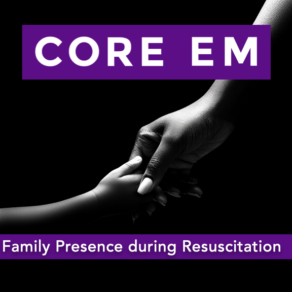 Episode 205: Family Presence during Resuscitation | We discuss the impact of family presence during resuscitations. Hosts: Ellen Duncan, MD, PhD Brian Gilberti, MD https://media.blubrry.com/coreem/content.blubrry.com/coreem/Family_Presence_During_Resuscitation.mp3 Download Leave a Comment Tags: Critical Care, Pediatrics Show Notes Overview Historical Context: The conversation around allowing family members in the room during resuscitation events began gaining attention in 1987. Since then, the practice has been increasingly encouraged. Current Practices in Pediatrics: Family presence during pediatric resuscitations remains inconsistent, with healthcare provider acceptance ranging from 15% to 85%. Many subspecialists and consultants still request that families step out, often due to outdated concerns. Common Concerns & Myths: Interference in resuscitation → Studies show minimal disruption. Legal risks → No increased litigation risk has been demonstrated. Family trauma → Research suggests that presence may help with grieving and reduce PTSD symptoms. Evidence from the Literature New England Journal of Medicine study on Family Presence During Cardiopulmonary Resuscitation (Jabre et al., 2013): In a randomized controlled trial of 570 relatives, PTSD-related symptoms were significantly higher in family members who were not offered the opportunity to be present during resuscitation. 79% of relatives in the intervention group witnessed CPR compared to 43% in the control group. Family members who did not witness CPR had a higher likelihood of PTSD symptoms (adjusted OR 1.7, p=0.004). Anxiety and depression symptoms were also higher in those who did not witness CPR. Impact on Medical Teams: The study found no evidence that family presence affected resuscitation success rates, medical team stress levels, or led to legal consequences. Health professionals’ concerns over interference were largely unfounded. Guideline Support & Barriers to Implementation Professional recommendations from pediatric societies support family presence during resuscitations. Barriers include: Lack of institutional policies ensuring family inclusion. Lack of formal training for providers on how to support families during these critical moments. Final Takeaways Encouraging institutional policy changes and training providers is key to implementing family presence during codes. Medical teams should challenge outdated practices and prioritize family-centered care in the emergency department. Family-witnessed resuscitation does not increase stress, legal risk, or compromise medical care—but it can significantly improve bereavement outcomes. Read More | — | ||||||
| 1/1/25 |  Episode 204: Necrotizing Fasciitis | We discuss the recognition and treatment of necrotizing fasciitis. Hosts: Aurnee Rahman, MD Brian Gilberti, MD https://media.blubrry.com/coreem/content.blubrry.com/coreem/Necrotizing_Fasciitis.mp3 Download Leave a Comment Tags: Critical Care, General Surgery Show Notes Table of Contents 0:00 – Introduction 0:41 – Overview 1:10 – Types of Necrotizing Fasciitis 2:21 – Pathophysiology & Risk Factors 3:16 – Clinical Presentation 4:06 – Diagnosis 5:37 – Treatment 7:09 – Prognosis and Recovery 7:37 – Take Home points Introduction Necrotizing soft tissue infections can be easily missed in routine cases of soft tissue infection. High mortality and morbidity underscore the need for vigilance. Definition A rapidly progressive, life-threatening infection of the deep soft tissues. Involves fascia and subcutaneous fat, causing fulminant tissue destruction. High mortality often due to delayed recognition and treatment. Types of Necrotizing Fasciitis Type I (Polymicrobial) Involves aerobic and anaerobic organisms (e.g., Bacteroides, Clostridium, Peptostreptococcus). Common in immunocompromised patients or those with comorbidities (e.g., diabetes, peripheral vascular disease). Type II (Monomicrobial) Often caused by Group A Streptococcus (Strep pyogenes) or Staphylococcus aureus. Can occur in otherwise healthy individuals. Vibrio vulnificus (associated with water exposure) is another example. Fournier’s Gangrene (Subset) Specific to perineal, genital, and perianal regions. Common in diabetic patients. Higher mortality, especially in females. Pathophysiology Spread Along Fascia Poor blood supply in fascial planes allows infection to advance rapidly. Tissue ischemia worsened by vascular thrombosis → rapid necrosis. High-Risk Patients Diabetes with vascular compromise. Recent surgeries or trauma (introducing bacteria into deep tissue). Immunosuppression (e.g., cirrhosis, malignancy, or immunosuppressive meds). NSAID use may mask symptoms, delaying diagnosis. Clinical Presentation Early Signs & Symptoms Severe Pain out of proportion to exam findings. Erythema (often with indistinct borders). Fever, Malaise (systemic signs of infection). Rapid progression with possible color changes (red → purple). Bullae Formation (fluid-filled blisters) and skin necrosis/gangrene. Crepitus in polymicrobial cases (gas production in tissue). Late-Stage Signs Systemic toxicity: hypotension, multi-organ failure if untreated. Diagnosis Clinical Suspicion Is Key Pain out of proportion, rapid progression, systemic signs. The “finger test” (small incision to explore fascial planes). Surgical Consultation Early surgical exploration is often the definitive diagnostic step. Lab Tests LRINEC Score (CRP, WBC, Hemoglobin, Sodium, Creatinine, Glucose) to stratify risk. Not definitive but can guide suspicion. Imaging CT scan may reveal gas in tissues, fascial edema, or muscle involvement. Must not delay surgical intervention if clinical suspicion is high. Treatment Principles Immediate & Aggressive Surgical Debridement Often multiple surgical procedures are required as necrosis progresses. Debridement back to healthy tissue margins. Empiric Broad-Spectrum Antibiotics Cover gram-positive (including MRSA), gram-negative, and anaerobes. Examples include: Vancomycin or Linezolid (for MRSA). Piperacillin-tazobactam or Carbapenems (for gram-negative & anaerobes). Clindamycin (to inhibit bacterial toxin production). Adjust based on culture results later. Adjunct Therapies Hyperbaric Oxygen Therapy (if available) for resistant cases. Evidence is mixed; not universally accessible. Supportive Care Intensive monitoring, often in an ICU setting. Fluid resuscitation & vasopressors for septic shock. Prognosis & Disposition High Mortality Rate Influenced by infection site, patient’s baseline health, and speed of intervention. Importance of Rapid Intervention Early recognition, aggressive surgery, and antibiotics improve survival. Long-Term Considerations Patients may require extensive rehabilitation. Reconstructive surgery often needed for tissue deficits. Disposition Operative management is mandatory; patients do not go home. Critical care admission is typical for hemodynamic monitoring and support. Five Key Take-Home Points High Suspicion Saves Lives: Recognize severe pain out of proportion as a critical red flag. Know Your NF Types & Risk Factors: Type I polymicrobial vs. Type II monomicrobial, plus subsets (Fournier’s). Clinical Diagnosis Above All: LRINEC and imaging help, but timely surgical exploration is paramount. Combined Surgical & Medical Therapy: Early debridement + broad-spectrum antibiotics (including toxin inhibition) is lifesaving. Extended Recovery & Mortality Risks: High mortality if missed or delayed. Expect prolonged rehab and possible multiple surgeries. Resources & Further Reading LRINEC Score Calculator EMCrit – Necrotizing Fasciitis Read More | — | ||||||
| 12/2/24 | 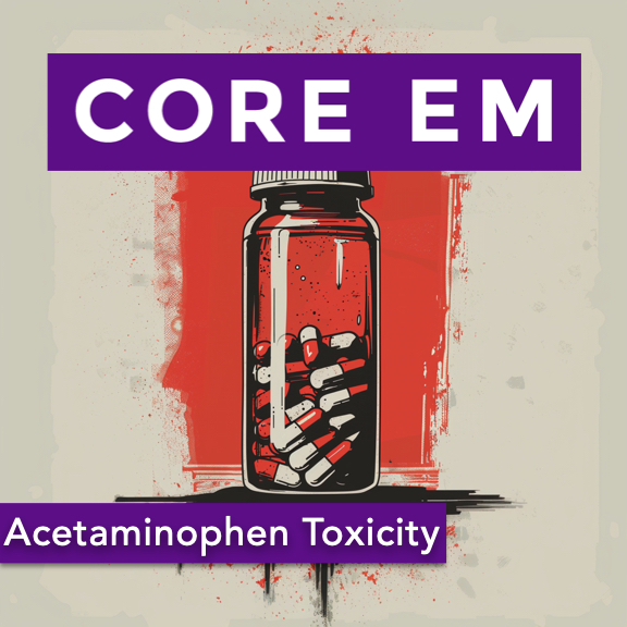 Episode 203: Acetaminophen Toxicity | We sit down with one of our toxicologists to discuss acetaminophen toxicity. Hosts: Marlis Gnirke, MD Brian Gilberti, MD https://media.blubrry.com/coreem/content.blubrry.com/coreem/Acetaminophen_Toxicity.mp3 Download One Comment Tags: Toxicology Show Notes Table of Contents 0:35 – Hidden acetaminophen toxicity in OTC products 3:24 – Pharmacokinetics and toxicokinetics 6:06 – Clinical Course 9:22 – The antidote – NAC 11:02 – The Rumack-Matthew Nomogram 17:36 – Treatment protocols 22:34 – Monitoring and Lab Work 23:23 – Considerations when treating pediatric patients 23:57 – IV APAP overdose, fomepizole 25:42 – Take Home Points Acetaminophen vs. Tylenol: The importance of recognizing that acetaminophen is found in many products beyond Tylenol. Common medications containing acetaminophen, such as Excedrin, Fioricet, Percocet, Dayquil/Nyquil, and others. The risk of unintentional overdose due to combination products. Prevalence of Acetaminophen Toxicity: Widespread availability and under-recognition contribute to its prevalence. The potential for unintentional overdose when taking multiple medications containing acetaminophen. Pharmacokinetics and Metabolism: Normal metabolism pathways of acetaminophen and the role of glutathione. Formation of the toxic metabolite NAPQI during overdose situations. Saturation of safe metabolic pathways leading to hepatotoxicity. Pathophysiology of Liver Injury: How excessive NAPQI leads to hepatocyte death, especially in zone III of the liver. The difference between therapeutic dosing and overdose metabolism. Clinical Stages of Acetaminophen Toxicity: Stage 1: Asymptomatic or nonspecific symptoms (first 24 hours). Stage 2: Onset of hepatic injury (24-72 hours), elevated AST/ALT. Stage 3: Maximum hepatotoxicity (72-96 hours), signs of liver failure. Stage 4: Recovery phase, complete hepatic regeneration if survived. Antidote – N-Acetylcysteine (NAC): Mechanisms of NAC in replenishing glutathione and detoxifying NAPQI. The importance of early administration, ideally within 8 hours post-ingestion. NAC’s role even in late presenters and in fulminant hepatic failure. The Rumack-Matthew Nomogram: How to use the nomogram for acute overdoses to determine the need for NAC. Limitations in chronic overdoses and late presentations. Emphasis on obtaining accurate time of ingestion and acetaminophen levels. Treatment Protocols: Standard 21-hour IV NAC protocol and dosing specifics. Managing anaphylactoid reactions associated with IV NAC. Criteria for extending NAC therapy beyond 21 hours. Monitoring and Laboratory Work: Importance of trending AST/ALT, INR, creatinine, lactate, and phosphate. Use of the King’s College Criteria for potential liver transplant evaluation. Special Considerations: Adjustments in pediatric patients regarding NAC dosing volumes. Awareness of IV acetaminophen overdoses and their management. Emerging discussions on the use of fomepizole in massive overdoses. Take-Home Points: Comprehensive Medication History: Always inquire about all medications taken to assess for potential acetaminophen exposure. Early Recognition and Treatment: Due to often silent initial stages, maintain a high index of suspicion and measure acetaminophen levels promptly. Understanding Metabolism and Toxicity: Recognize how overdose alters metabolism, leading to toxic NAPQI accumulation. N-Acetylcysteine Efficacy: NAC is most effective when administered early but remains beneficial even in advanced stages. Proper Use of the Nomogram: Utilize the Rumack-Matthew Nomogram appropriately for acute ingestions and consult toxicology when in doubt. Monitoring and Continuing Care: Be vigilant in monitoring laboratory values and prepared to extend NAC therapy as needed. Consultation and Resources: Engage with poison control centers and utilize available resources for complex cases. Resources Mentioned Rumack-Matthew Nomogram Rumack-Matthew Nomogram, credit: MDCalc King’s College Criteria King’s College Criteria for Acetaminophen Toxicity Use this tool to assess the need for liver transplant evaluation in cases of acetaminophen-induced hepatic failure. Includes criteria for pH, INR, creatinine, and more. Poison Control Center (available 24/7 for consultation): 1-800-222-1222 References Goldfrank’s Toxicologic Emergencies, 9th Edition was consulted for information on the pharmacokinetics and clinical presentation of acetaminophen toxicity. For more details, see: Nelson, L. S., Howland, M. A., Lewin, N. A., Smith, S. W., Goldfrank, L. R., & Hoffman, R. S. (Eds.). (2011). Goldfrank’s toxicologic emergencies (9th ed.). McGraw-Hill Education. Read More | — | ||||||
| 11/1/24 | 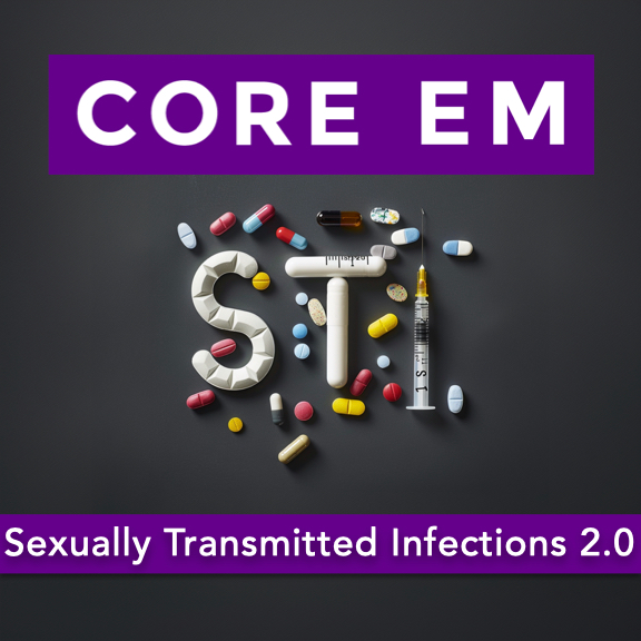 Episode 202: Sexually Transmitted Infections 2.0 | We review Sexually Transmitted Infections and pertinent updates in diagnosis and management. Hosts: Avir Mitra, MD Brian Gilberti, MD https://media.blubrry.com/coreem/content.blubrry.com/coreem/Sexually_Transmitted_Infections_2_0.mp3 Download Leave a Comment Tags: gynecology, Infectious Diseases, Urology Show Notes Table of Contents (1:49) Chlamydia (3:31) Gonorrhea (4:50) PID (6:14) Syphilis (8:08) Neurosyphilis (9:13) Tertiary Syphilis (10:06) Trichomoniasis (11:13) Herpes (12:49) HIV (14:10) PEP (15:13) Mycoplasma Genitalium (18:00) Take Home Points Chlamydia: Prevalence: Most common STI. High percentage of asymptomatic cases (40% to 96%). Presentation: Urethritis, cervicitis, pelvic inflammatory disease (PID), prostatitis, proctitis, pharyngitis, arthritis. Importance of considering extra-genital sites (oral and rectal infections). Testing: Gold Standard: Nucleic Acid Amplification Test (NAAT) via PCR. Sampling Sites: Endocervical or urethral swabs preferred over urine samples due to higher sensitivity. Triple-site testing (genital, rectal, pharyngeal) recommended for comprehensive detection. Treatment Updates: Previous Regimen: Azithromycin 1 g orally in a single dose. Current First-Line Treatment: Doxycycline 100 mg orally twice daily for 7 days. Alternatives: Azithromycin remains an option for patients unlikely to adhere to a 7-day regimen or for pregnant patients. Note: PID treatment differs and will be discussed separately. Gonorrhea: Presentation: Similar to chlamydia; can be asymptomatic. Symptoms include urethritis, cervicitis, PID, prostatitis, proctitis, pharyngitis. Testing: Gold Standard: NAAT. Sampling Sites: Endocervical swabs are more sensitive than urine samples. Triple-site testing is crucial to avoid missing infections. Treatment Updates: Previous Regimen: Ceftriaxone 250 mg IM plus azithromycin 1 g orally. Current Recommendation: Ceftriaxone 500 mg IM single dose. Adjusted due to rising azithromycin resistance and updated pharmacokinetic data. Co-Infection Considerations: High rates of chlamydia and gonorrhea co-infection (20% to 40%). CDC recommends empiric treatment for chlamydia when treating gonorrhea to prevent complications like PID and infertility. Pelvic Inflammatory Disease (PID): Etiology: Not solely caused by chlamydia and gonorrhea; about 50% of cases involve other pathogens like bacterial vaginosis (BV) organisms and anaerobes. Treatment Changes: Expanded Coverage Regimen: Ceftriaxone 500 mg IM once. Doxycycline 100 mg orally twice daily for 14 days. Metronidazole 500 mg orally twice daily for 14 days. Inclusion of metronidazole addresses anaerobic bacteria contributing to PID. Syphilis: Stages and Presentation: Primary Syphilis: Painless chancre on genitals. Treatment: Penicillin G 2.4 million units IM single dose. Secondary Syphilis: Rash (often diffuse), mucocutaneous lesions, nonspecific joint pain. Treatment: Same as primary syphilis. Latent Syphilis: Asymptomatic phase; divided into early (<1 year) and late (>1 year). Treatment for Late Latent: Penicillin G 2.4 million units IM once weekly for 3 weeks. Recommended when the timing of infection is unclear. Neurosyphilis: Can occur at any stage. Symptoms include visual changes, severe headaches, neurological deficits. Diagnosis: Requires lumbar puncture (LP) for confirmation. Treatment: Admission for intravenous penicillin G. Tertiary Syphilis: Rare, advanced stage with severe manifestations (e.g., gummas, cardiovascular complications, neurological signs). Treatment: Extended penicillin therapy similar to late latent syphilis. Trichomoniasis: Presentation: Often asymptomatic. In women: Vaginal discharge. In men: Urethritis. Testing: Shift from wet mount microscopy to NAAT for improved detection. Swab samples preferred over urine for higher sensitivity. Treatment Updates: Previous Regimen: Metronidazole 2 g orally in a single dose. Current Recommendations: Women: Metronidazole 500 mg orally twice daily for 7 days. Men: Single 2 g dose remains acceptable. Herpes Simplex Virus (HSV): Types and Transmission: HSV-1 and HSV-2: Both can cause oral and genital infections. Increasing crossover between oral and genital sites. Testing: Serum IgG testing not useful for acute diagnosis due to widespread prior exposure. Preferred Method: PCR testing from lesion swabs. Clinical Tip: If the lesion is characteristic, clinicians may start treatment without waiting for test results. Treatment: Preferred Medication: Valacyclovir (Valtrex) for ease of dosing. Dosage: Initial episode: 1 g orally twice daily for 7 to 10 days. Recurrence: 1 g daily for 5 days. Alternative: Acyclovir for cost considerations. Human Immunodeficiency Virus (HIV): Testing Limitations: Window Periods: Fourth-generation tests have a window period of 2 to 4 weeks. Negative results during this period may not rule out recent infection. Acute HIV Infection: Presents with flu-like symptoms: malaise, joint pains, fatigue. Diagnosis Challenges: Standard HIV tests may be negative during the window period. Options: Empiric treatment with follow-up testing. Order an HIV viral load test (more sensitive but expensive and delayed results). Post-Exposure Prophylaxis (PEP): Timing: Initiate ideally within 72 hours of potential exposure. Duration: 28-day regimen. Pre-Treatment Testing: Baseline HIV test to rule out existing infection. Renal and hepatic function tests to monitor for medication side effects. Follow-Up: Reassess renal/hepatic function in 2 weeks. Mycoplasma genitalium: Recognition: Newly recognized STI by the CDC in 2021. Causes cervicitis and urethritis. Possible associations with PID and proctitis, but not definitively established. Testing: When to Test: Only in patients with persistent symptoms after standard STI testing and treatment. Not recommended for initial screening. Method: NAAT. Treatment: Step 1: Doxycycline 100 mg orally twice daily for 7 days. Step 2: Moxifloxacin 400 mg orally once daily for 7 days. Addresses antibiotic resistance concerns and ensures comprehensive treatment. General Management and Patient Counseling: Partner Notification: Encourage patients to inform sexual partners for testing and treatment. Medication Adherence: Emphasize the importance of completing the full course of prescribed medications. Prevention Measures: Discuss the use of barrier protection (e.g., condoms) to prevent transmission and reinfection. Follow-Up Care: Advise patients to return if symptoms persist, indicating possible infections like Mycoplasma genitalium. Key Take-Home Points: Chlamydia Treatment Update: Doxycycline 100 mg orally twice daily for 7 days is now first-line treatment for cervical infections. For epididymitis, extend doxycycline to 10 days. Gonorrhea Treatment Update: Treat with a single 500 mg IM dose of ceftriaxone. PID Management Update: Expanded antimicrobial coverage includes: Ceftriaxone 500 mg IM once. Doxycycline 100 mg orally twice daily for 14 days. Metronidazole 500 mg orally twice daily for 14 days. Mycoplasma genitalium Recognition: Test in patients with persistent symptoms after standard treatment. Treat with doxycycline followed by moxifloxacin. HIV Testing and PEP: Be aware of HIV test window periods; negative results may not rule out recent infection. Consider HIV viral load testing if acute infection is suspected. Initiate PEP within 72 hours for a 28-day course, ensuring clear discharge planning and patient support. Read More | — | ||||||
| 10/1/24 |  Episode 201: Migraines | We discuss migraines with one of the authorities in the field. Hosts: Benjamin Friedman, MD of Montefiore Brian Gilberti, MD https://media.blubrry.com/coreem/content.blubrry.com/coreem/Migraines.mp3 Download Leave a Comment Tags: Neurology Show Notes Initial Approach to Diagnosing Migraines: Differentiating between primary headaches (migraine, tension-type, cluster) and secondary causes (e.g., subarachnoid hemorrhage). The importance of patient history and reevaluation after initial treatment. Recognizing the unique presentation of cluster headaches and their management implications. Effective Acute Migraine Treatments: First-line treatments including anti-dopaminergic medications like metoclopramide (Reglan) and prochlorperazine (Compazine), and parenteral NSAIDs like ketorolac (Toradol). The limited role of triptans in the ED due to side effects and less efficacy compared to anti-dopaminergics. The use of nerve blocks (greater occipital nerve block and sphenopalatine ganglion block) as effective treatments without systemic side effects. Treatments to Avoid or Use with Caution: Diphenhydramine (Benadryl): Studies show it does not prevent akathisia from anti-dopaminergics nor improve migraine outcomes. IV Fluids: Routine use is not supported unless the patient shows signs of dehydration. Magnesium: Conflicting evidence with some studies showing no benefit or even harm. Managing Refractory Migraines: Second-line treatments including additional doses of metoclopramide combined with NSAIDs or dihydroergotamine (DHE). Considering opioids as a last resort when other treatments fail. The potential use of newer medications like lasmiditan and CGRP antagonists. Preventing Recurrence of Migraines: Administering a single dose of dexamethasone (4 mg IV) to reduce the risk of headache recurrence after discharge. Prescribing NSAIDs or triptans upon discharge for outpatient management. Recognizing and addressing chronic migraine, and initiating preventive therapies like propranolol when appropriate. Key Takeaways Differentiate Primary from Secondary Headaches and Reassess After Treatment: Use patient history and reevaluation post-treatment to distinguish migraines from more serious conditions, reducing unnecessary imaging and procedures. First-Line Treatments Are Effective: Anti-dopaminergic medications and NSAIDs are the mainstay of acute migraine treatment in the ED. Reserve opioids for cases unresponsive to multiple lines of treatment. Avoid Unnecessary Interventions: Diphenhydramine and routine IV fluids do not have proven benefits and can be excluded to streamline care. Utilize Nerve Blocks for Refractory Cases: Greater occipital nerve blocks and sphenopalatine ganglion blocks are effective alternatives for patients not responding to medication. Prevent Recurrence with Dexamethasone and Outpatient Planning: A single IV dose of dexamethasone can help prevent recurrence. Provide prescriptions and consider preventive therapies to reduce future ED visits. Read More | — | ||||||
| 9/2/24 | Episode 200: Immune Checkpoint Inhibitors | We discuss a new class of medications, Immune Checkpoint Inhibitors, and their side effects. Hosts: Avir Mitra, MD Brian Gilberti, MD https://media.blubrry.com/coreem/content.blubrry.com/coreem/Immune_Checkpoint_Inhibitors.mp3 Download Leave a Comment Tags: Oncology Show Notes Overview of Immune Checkpoint Inhibitors (ICIs) ICIs are a relatively new class of oncologic drugs that have revolutionized cancer treatment. Unlike chemotherapy, ICIs help the immune system develop memory against cancer cells and adapt as the cancer mutates. Since their release in 2011, ICIs have expanded to 83 indications for 17 different cancers, with approximately 230,000 patients using them. Mechanism of Action Cancer cells can evade the immune system by binding to T cell receptors that downregulate the immune response. ICIs work by blocking these receptors or ligands, preventing the downregulation and allowing T cells to proliferate and attack cancer cells. Common ICIs Risks and Toxicities of ICIs ICIs can lead to autoimmune attacks on healthy cells due to immune system upregulation. Immune-related adverse effects (irAEs) include colitis, pneumonitis, dermatitis, hepatitis, and endocrine issues (e.g., hypothyroid, hypocortisolemia, hypophysitis). These toxicities can present as infections, making diagnosis challenging in the emergency room. Management of ICI Toxicities in the ER Diagnosis: Look for signs that mimic infections (e.g., cough and fever in pneumonitis). Diagnostic Imaging in pneumonitis: If CXR is normal but suspicion is high, consider CT scans to differentiate conditions like pneumonitis from other issues such as malignancy-associated pleural effusion or acute pulmonary embolism. Treatment: The primary treatment for irAEs is steroids (e.g., prednisone 1 mg/kg). Start steroids early and hold the ICI to manage symptoms effectively and increase the likelihood of resuming ICI therapy later. Consider using antibiotics in combination with steroids if there is uncertainty about whether symptoms are due to infection or ICI toxicity. Coordinate care with the patient’s oncologist if possible Disposition Decisions Patient disposition (admit vs. discharge) should depend on clinical presentation and severity. Coordination with oncology is crucial; they are often comfortable with starting steroids even if there is a potential infection. Patients can be discharged if symptoms are mild, but sicker patients with more complex presentations may require admission. Take-Home Points ICIs are a new class of cancer drugs that effectively target cancer cells but come with unique immune-related toxicities. Diagnosing irAEs can be challenging due to symptom overlap with infections. The cornerstone of treatment is early administration of steroids and temporarily holding the ICI. Close collaboration with oncology teams is essential for optimal patient management. Read More | — | ||||||
Showing 25 of 72
Sponsor Intelligence
Sign in to see which brands sponsor this podcast, their ad offers, and promo codes.
Chart Positions
36 placements across 30 markets.
Chart Positions
36 placements across 30 markets.